

Anti-Laminin [A5]
Ab02770-10.0
ApplicationsImmunoFluorescence, ImmunoPrecipitation, ImmunoHistoChemistry
Product group Antibodies
ReactivityBovine, Human, Mouse
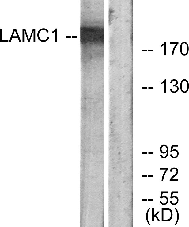
TargetLAMC1
Overview
- SupplierAbsolute Antibody
- Product NameAnti-Laminin [A5]
- Delivery Days Customer7
- Application Supplier NoteThis original rat IgG2a clone can be used to detect and analyze the presence of laminin. The specificity towards this protein has been confirmed using immunoprecipitation (Ljubimov et al., 1986; PMID: 3522258). The location of the laminin was confirmed to be in the basment membrane by means of immunofluorescence using this antibody (Eriksdotter-Nielson et al., 1986; PMID: 3537540).
- ApplicationsImmunoFluorescence, ImmunoPrecipitation, ImmunoHistoChemistry
- Applications SupplierIP; IHC; IF
- CertificationResearch Use Only
- ClonalityMonoclonal
- Clone IDA5
- Gene ID3915
- Target nameLAMC1
- Target descriptionlaminin subunit gamma 1
- Target synonymsLAMB2, laminin subunit gamma-1, S-LAM gamma, S-laminin subunit gamma, laminin B2 chain, laminin, gamma 1 (formerly LAMB2), laminin-10 subunit gamma, laminin-11 subunit gamma, laminin-2 subunit gamma, laminin-3 subunit gamma, laminin-4 subunit gamma, laminin-6 subunit gamma, laminin-7 subunit gamma, laminin-8 subunit gamma, laminin-9 subunit gamma
- HostHuman
- IsotypeIgG1
- Protein IDP11047
- Protein NameLaminin subunit gamma-1
- Scientific DescriptionThis chimeric human antibody was made using the variable domain sequences of the original Rat IgG2a format, for improved compatibility with existing reagents, assays and techniques.
- ReactivityBovine, Human, Mouse
- Reactivity SupplierHuman, Mouse, Bovine
- Reactivity Supplier NoteThe original rat IgG2a version of this antibody was raised by immunizing fisher rats with high molecular mass material from the Engelbreth-Holm-Swarm (EHS) tumor matrix containing laminin, entactin and HSPG, and fusing the spleen cells of these rats with mouse myeloma X63 Ag8.653 cells.
- Storage Instruction-20°C,2°C to 8°C
- UNSPSC41116161
Related products
Product group Antibodies
Anti-LAMC1 AntibodyA97431
ApplicationsWestern Blot, ELISA
ReactivityHuman, Mouse, Rat
- SizePrice

Product group Antibodies
Anti-Laminin gamma 1/LAMC1 Antibody Picoband(r)A03522-2-CARRIER-FREE
ApplicationsWestern Blot, ELISA
ReactivityHuman, Mouse, Rat
TargetLAMC1
- SizePrice
Product group Antibodies
Anti-LAMC1 Antibody144-64027
ApplicationsImmunoFluorescence, Western Blot, ImmunoHistoChemistry
ReactivityHuman, Mouse, Rat
TargetLAMC1
- SizePrice

Product group Antibodies
Anti-LAMC1 AntibodyAMAB91136
ApplicationsWestern Blot, ImmunoHistoChemistry
ReactivityHuman
TargetLAMC1
- SizePrice
Product group Antibodies
ApplicationsFlow Cytometry, Western Blot, ImmunoCytoChemistry
ReactivityHuman, Rat
TargetLAMC1
- SizePrice
Product group Antibodies
LAMC1 AntibodyCSB-PA009776
ApplicationsImmunoFluorescence, Western Blot, ELISA, ImmunoHistoChemistry
ReactivityHuman, Monkey, Mouse, Rat
TargetLAMC1
- SizePrice
Product group Antibodies
ApplicationsImmunoPrecipitation, Western Blot, ImmunoCytoChemistry, ImmunoHistoChemistry
ReactivityMouse, Rat
TargetLAMC1
- SizePrice
Product group Antibodies

Rat anti LamininMUB1100P
ApplicationsImmunoFluorescence, ImmunoCytoChemistry, ImmunoHistoChemistry, ImmunoHistoChemistry Frozen
ReactivityHuman, Mouse, Porcine
TargetLAMC1
- SizePrice
Product group Antibodies
ApplicationsELISA, ImmunoHistoChemistry
ReactivityHuman
TargetLAMC1
- SizePrice
Product group Antibodies

ApplicationsWestern Blot, ELISA, ImmunoHistoChemistry, ImmunoHistoChemistry Paraffin
ReactivityHuman
TargetLAMC1
- SizePrice