


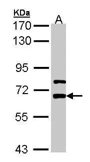
Figure 1. Western blot analysis of Lgi1/EPT/LGI1 using anti-Lgi1/EPT/LGI1 antibody (A00850-1). Electrophoresis was performed on a 5-20% SDS-PAGE gel at 70V (Stacking gel) / 90V (Resolving gel) for 2-3 hours. The sample well of each lane was loaded with 50ug of sample under reducing conditions. Lane 1: human SH-SY5Y whole cell lysates, Lane 2: rat C6 whole cell lysates, Lane 3: mouse Neuro-2a whole cell lysates, Lane 4: human HEK293 whole cell lysates. After Electrophoresis, proteins were transferred to a Nitrocellulose membrane at 150mA for 50-90 minutes. Blocked the membrane with 5% Non-fat Milk/ TBS for 1.5 hour at RT. The membrane was incubated with rabbit anti-Lgi1/EPT/LGI1 antigen affinity purified polyclonal antibody (Catalog # A00850-1) at 0.5 microg/mL overnight at 4°C, then washed with TBS-0.1%Tween 3 times with 5 minutes each and probed with a goat anti-rabbit IgG-HRP secondary antibody at a dilution of 1:5000 for 1.5 hour at RT. The signal is developed using an Enhanced Chemiluminescent detection (ECL) kit (Catalog # EK1002) with Tanon 5200 system. A specific band was detected for Lgi1/EPT/LGI1 at approximately 64KD. The expected band size for Lgi1/EPT/LGI1 is at 64KD.
. Overlay histogram showing U20S cells stained with A00850-1 (Blue line). The cells were fixed with 4% paraformaldehyde and blocked with 10% normal goat serum. And then incubated with rabbit anti-Lgi1/EPT/LGI1 Antibody (A00850-1, 1microg/1x106 cells) for 30 min at 20°C. DyLight®488 conjugated goat anti-rabbit IgG (BA1127, 5-10microg/1x106 cells) was used as secondary antibody for 30 minutes at 20°C. Isotype control antibody (Green line) was rabbit IgG (1microg/1x106) used under the same conditions. Unlabelled sample without incubation with primary antibody and secondary antibody (Red line) was used as a blank control.")
Figure 1. Western blot analysis of Lgi1/EPT/LGI1 using anti-Lgi1/EPT/LGI1 antibody (A00850-1). Electrophoresis was performed on a 5-20% SDS-PAGE gel at 70V (Stacking gel) / 90V (Resolving gel) for 2-3 hours. The sample well of each lane was loaded with 50ug of sample under reducing conditions. Lane 1: human SH-SY5Y whole cell lysates, Lane 2: rat C6 whole cell lysates, Lane 3: mouse Neuro-2a whole cell lysates, Lane 4: human HEK293 whole cell lysates. After Electrophoresis, proteins were transferred to a Nitrocellulose membrane at 150mA for 50-90 minutes. Blocked the membrane with 5% Non-fat Milk/ TBS for 1.5 hour at RT. The membrane was incubated with rabbit anti-Lgi1/EPT/LGI1 antigen affinity purified polyclonal antibody (Catalog # A00850-1) at 0.5 microg/mL overnight at 4°C, then washed with TBS-0.1%Tween 3 times with 5 minutes each and probed with a goat anti-rabbit IgG-HRP secondary antibody at a dilution of 1:5000 for 1.5 hour at RT. The signal is developed using an Enhanced Chemiluminescent detection (ECL) kit (Catalog # EK1002) with Tanon 5200 system. A specific band was detected for Lgi1/EPT/LGI1 at approximately 64KD. The expected band size for Lgi1/EPT/LGI1 is at 64KD.
Anti-Lgi1/EPT/LGI1 Antibody Picoband(r)
A00850-1-CARRIER-FREE
ApplicationsFlow Cytometry, Western Blot, ELISA
Product group Antibodies
ReactivityHuman, Mouse, Rat
TargetLGI1
Overview
- SupplierBoster Bio
- Product NameAnti-Lgi1/EPT/LGI1 Antibody Picoband(r)
- Delivery Days Customer9
- ApplicationsFlow Cytometry, Western Blot, ELISA
- CertificationResearch Use Only
- ClonalityPolyclonal
- Concentration500 ug/ml
- Gene ID9211
- Target nameLGI1
- Target descriptionleucine rich glioma inactivated 1
- Target synonymsADLTE, ADPAEF, ADPEAF, EPITEMPIN, EPT, ETL1, IB1099, leucine-rich glioma-inactivated protein 1, epitempin-1
- HostRabbit
- IsotypeIgG
- Protein IDO95970
- Protein NameLeucine-rich glioma-inactivated protein 1
- Scientific DescriptionBoster Bio Anti-Lgi1/EPT/LGI1 Antibody Picoband® catalog # A00850-1. Tested in ELISA, Flow Cytometry, WB applications. This antibody reacts with Human, Mouse, Rat. The brand Picoband indicates this is a premium antibody that guarantees superior quality, high affinity, and strong signals with minimal background in Western blot applications. Only our best-performing antibodies are designated as Picoband, ensuring unmatched performance.
- ReactivityHuman, Mouse, Rat
- Storage Instruction-20°C,2°C to 8°C
- UNSPSC12352203
Related products
Product group Antibodies
LGI1 AntibodyCSB-PA012898ESR2HU
ApplicationsELISA, ImmunoHistoChemistry
ReactivityHuman
TargetLGI1
- SizePrice
Product group Antibodies
Anti-LGI1 AntibodyA48397
ApplicationsWestern Blot, ELISA, ImmunoHistoChemistry
ReactivityHuman, Mouse, Rat
- SizePrice
Product group Antibodies

LGI1 Antibody (aa35-285)LS-C376024
ApplicationsELISA, ImmunoHistoChemistry, ImmunoHistoChemistry Paraffin
ReactivityHuman
TargetLGI1
- SizePrice
Product group Antibodies
LGI1 antibody [N2C2], InternalGTX105700
ApplicationsImmunoFluorescence, Western Blot, ImmunoCytoChemistry
ReactivityHuman, Rat
TargetLGI1
- SizePrice
Product group Antibodies
ApplicationsFlow Cytometry, Western Blot, ImmunoCytoChemistry
ReactivityHuman
TargetLGI1
- SizePrice
Product group Antibodies
Anti-LGI1 Antibody144-61416
ApplicationsWestern Blot, ImmunoHistoChemistry
ReactivityHuman, Mouse, Rat
TargetLGI1
- SizePrice