

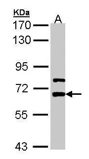
Sample (30 ug of whole cell lysate) A: A431 (GTX27909) 7.5% SDS PAGE GTX105700 diluted at 1:5000
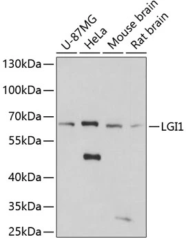
![Various whole cell extracts (30 μg) were separated by 7.5% SDS-PAGE, and the membrane was blotted with LGI1 antibody [N2C2], Internal (GTX105700) diluted at 1:1000. The HRP-conjugated anti-rabbit IgG antibody (GTX213110-01) was used to detect the primary antibody.](https://www.genetex.com/upload/website/prouct_img/normal/GTX105700/GTX105700_40030_20180316_WB_w_23060120_663.webp "Various whole cell extracts (30 μg) were separated by 7.5% SDS-PAGE, and the membrane was blotted with LGI1 antibody [N2C2], Internal (GTX105700) diluted at 1:1000. The HRP-conjugated anti-rabbit IgG antibody (GTX213110-01) was used to detect the primary antibody.")
![LGI1 antibody [N2C2], Internal detects LGI1 protein by immunofluorescent analysis. Sample: DIV10 rat E18 primary hippocampal neurons were fixed in 4% paraformaldehyde at RT for 15 min. Green: LGI1 protein stained by LGI1 antibody [N2C2], Internal (GTX105700) diluted at 1:500. Red: beta Tubulin 3/ Tuj1, stained by beta Tubulin 3/ Tuj1 antibody [GT1338] (GTX631831) diluted at 1:500. Blue: Fluoroshield with DAPI (GTX30920).](https://www.genetex.com/upload/website/prouct_img/normal/GTX105700/GTX105700_40030_20170824_IFA_w_23060120_528.webp "LGI1 antibody [N2C2], Internal detects LGI1 protein by immunofluorescent analysis. Sample: DIV10 rat E18 primary hippocampal neurons were fixed in 4% paraformaldehyde at RT for 15 min. Green: LGI1 protein stained by LGI1 antibody [N2C2], Internal (GTX105700) diluted at 1:500. Red: beta Tubulin 3/ Tuj1, stained by beta Tubulin 3/ Tuj1 antibody [GT1338] (GTX631831) diluted at 1:500. Blue: Fluoroshield with DAPI (GTX30920).")
![Rat tissue extract (50 μg) was separated by 7.5% SDS-PAGE, and the membrane was blotted with LGI1 antibody [N2C2], Internal (GTX105700) diluted at 1:1000. The HRP-conjugated anti-rabbit IgG antibody (GTX213110-01) was used to detect the primary antibody.](https://www.genetex.com/upload/website/prouct_img/normal/GTX105700/GTX105700_40030_20180316_WB_R_brain_w_23060120_430.webp "Rat tissue extract (50 μg) was separated by 7.5% SDS-PAGE, and the membrane was blotted with LGI1 antibody [N2C2], Internal (GTX105700) diluted at 1:1000. The HRP-conjugated anti-rabbit IgG antibody (GTX213110-01) was used to detect the primary antibody.")
Sample (30 ug of whole cell lysate) A: A431 (GTX27909) 7.5% SDS PAGE GTX105700 diluted at 1:5000
LGI1 antibody [N2C2], Internal
GTX105700
ApplicationsImmunoFluorescence, Western Blot, ImmunoCytoChemistry
Product group Antibodies
ReactivityHuman, Rat
TargetLGI1
Overview
- SupplierGeneTex
- Product NameLGI1 antibody [N2C2], Internal
- Delivery Days Customer9
- Application Supplier NoteWB: 1:500-1:10000. ICC/IF: 1:100-1:1000. *Optimal dilutions/concentrations should be determined by the researcher.Not tested in other applications.
- ApplicationsImmunoFluorescence, Western Blot, ImmunoCytoChemistry
- CertificationResearch Use Only
- ClonalityPolyclonal
- Concentration1 mg/ml
- ConjugateUnconjugated
- Gene ID9211
- Target nameLGI1
- Target descriptionleucine rich glioma inactivated 1
- Target synonymsADLTE, ADPAEF, ADPEAF, EPITEMPIN, EPT, ETL1, IB1099, leucine-rich glioma-inactivated protein 1, epitempin-1
- HostRabbit
- IsotypeIgG
- Protein IDO95970
- Protein NameLeucine-rich glioma-inactivated protein 1
- Scientific DescriptionThis gene is rearranged as a result of translocations in glioblastoma cell lines. The protein contains a hydrophobic segment representing a putative transmembrane domain with the amino terminus located outside the cell. It also contains leucine-rich repeats with conserved cysteine-rich flanking sequences. This gene is predominantly expressed in neural tissues and its expression is reduced in low grade brain tumors and significantly reduced or absent in malignant gliomas. Mutations in this gene result in autosomal dominant lateral temporal epilepsy. [provided by RefSeq]
- ReactivityHuman, Rat
- Storage Instruction-20°C or -80°C,2°C to 8°C
- UNSPSC41116161
Datasheet
Related products
Product group Antibodies
LGI1 AntibodyCSB-PA012898ESR2HU
ApplicationsELISA, ImmunoHistoChemistry
ReactivityHuman
TargetLGI1
- SizePrice

Product group Antibodies
Anti-Lgi1/EPT/LGI1 Antibody Picoband(r)A00850-1-CARRIER-FREE
ApplicationsFlow Cytometry, Western Blot, ELISA
ReactivityHuman, Mouse, Rat
TargetLGI1
- SizePrice
Product group Antibodies
Anti-LGI1 AntibodyA48397
ApplicationsWestern Blot, ELISA, ImmunoHistoChemistry
ReactivityHuman, Mouse, Rat
- SizePrice
Product group Antibodies

LGI1 Antibody (aa35-285)LS-C376024
ApplicationsELISA, ImmunoHistoChemistry, ImmunoHistoChemistry Paraffin
ReactivityHuman
TargetLGI1
- SizePrice
Product group Antibodies
LGI1 antibodyGTX66465
ApplicationsWestern Blot
ReactivityHuman, Mouse, Rat
TargetLGI1
- SizePrice
Product group Antibodies
ApplicationsFlow Cytometry, Western Blot, ImmunoCytoChemistry
ReactivityHuman
TargetLGI1
- SizePrice
Product group Antibodies
Anti-LGI1 Antibody144-61416
ApplicationsWestern Blot, ImmunoHistoChemistry
ReactivityHuman, Mouse, Rat
TargetLGI1
- SizePrice