


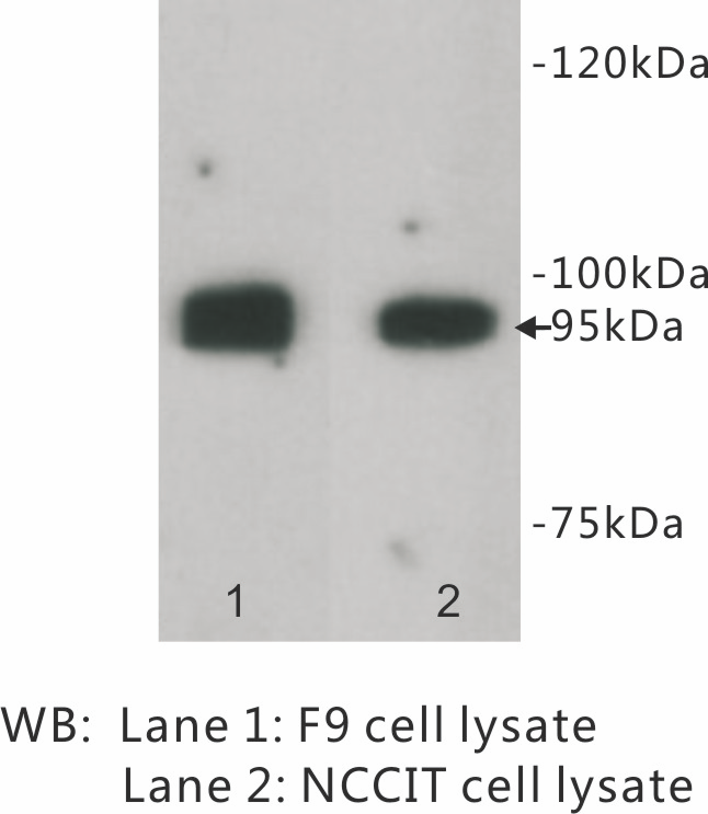
Figure 1. Western blot analysis of LGR5 using anti-LGR5 antibody (A00239-2). Electrophoresis was performed on a 5-20% SDS-PAGE gel at 70V (Stacking gel) / 90V (Resolving gel) for 2-3 hours. The sample well of each lane was loaded with 30 ug of sample under reducing conditions. Lane 1: human SH-SY5Y whole cell lysates, Lane 2: human HepG2 whole cell lysates, Lane 3: human RT4 whole cell lysates, Lane 4: human 293T whole cell lysates. After electrophoresis, proteins were transferred to a nitrocellulose membrane at 150 mA for 50-90 minutes. Blocked the membrane with 5% non-fat milk/TBS for 1.5 hour at RT. The membrane was incubated with rabbit anti-LGR5 antigen affinity purified polyclonal antibody (Catalog # A00239-2) at 0.5 microg/mL overnight at 4°C, then washed with TBS-0.1%Tween 3 times with 5 minutes each and probed with a goat anti-rabbit IgG-HRP secondary antibody at a dilution of 1:5000 for 1.5 hour at RT. The signal is developed using an Enhanced Chemiluminescent detection (ECL) kit (Catalog # EK1002) with Tanon 5200 system. A specific band was detected for LGR5 at approximately 130 kDa. The expected band size for LGR5 is at 100,97,92 kDa.
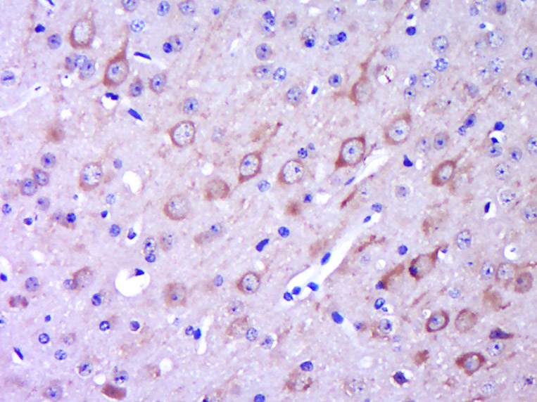
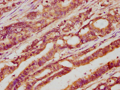
. LGR5 was detected in a paraffin-embedded section of mouse small intestine tissue. Heat mediated antigen retrieval was performed in EDTA buffer (pH 8.0, epitope retrieval solution). The tissue section was blocked with 10% goat serum. The tissue section was then incubated with 2 microg/ml rabbit anti-LGR5 Antibody (A00239-2) overnight at 4°C. Peroxidase Conjugated Goat Anti-rabbit IgG was used as secondary antibody and incubated for 30 minutes at 37°C. The tissue section was developed using HRP Conjugated Rabbit IgG Super Vision Assay Kit (Catalog # SV0002) with DAB as the chromogen.")
. LGR5 was detected in a paraffin-embedded section of rat small intestine tissue. Heat mediated antigen retrieval was performed in EDTA buffer (pH 8.0, epitope retrieval solution). The tissue section was blocked with 10% goat serum. The tissue section was then incubated with 2 microg/ml rabbit anti-LGR5 Antibody (A00239-2) overnight at 4°C. Peroxidase Conjugated Goat Anti-rabbit IgG was used as secondary antibody and incubated for 30 minutes at 37°C. The tissue section was developed using HRP Conjugated Rabbit IgG Super Vision Assay Kit (Catalog # SV0002) with DAB as the chromogen.")
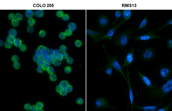
. Overlay histogram showing 293T cells stained with A00239-2 (Blue line). The cells were fixed with 4% paraformaldehyde and blocked with 10% normal goat serum. And then incubated with rabbit anti-LGR5 Antibody (A00239-2, 1 microg/1x106 cells) for 30 min at 20°C. DyLight®488 conjugated goat anti-rabbit IgG (BA1127, 5-10 microg/1x106 cells) was used as secondary antibody for 30 minutes at 20°C. Isotype control antibody (Green line) was rabbit IgG (1 microg/1x106) used under the same conditions. Unlabelled sample without incubation with primary antibody and secondary antibody (Red line) was used as a blank control.")
Figure 1. Western blot analysis of LGR5 using anti-LGR5 antibody (A00239-2). Electrophoresis was performed on a 5-20% SDS-PAGE gel at 70V (Stacking gel) / 90V (Resolving gel) for 2-3 hours. The sample well of each lane was loaded with 30 ug of sample under reducing conditions. Lane 1: human SH-SY5Y whole cell lysates, Lane 2: human HepG2 whole cell lysates, Lane 3: human RT4 whole cell lysates, Lane 4: human 293T whole cell lysates. After electrophoresis, proteins were transferred to a nitrocellulose membrane at 150 mA for 50-90 minutes. Blocked the membrane with 5% non-fat milk/TBS for 1.5 hour at RT. The membrane was incubated with rabbit anti-LGR5 antigen affinity purified polyclonal antibody (Catalog # A00239-2) at 0.5 microg/mL overnight at 4°C, then washed with TBS-0.1%Tween 3 times with 5 minutes each and probed with a goat anti-rabbit IgG-HRP secondary antibody at a dilution of 1:5000 for 1.5 hour at RT. The signal is developed using an Enhanced Chemiluminescent detection (ECL) kit (Catalog # EK1002) with Tanon 5200 system. A specific band was detected for LGR5 at approximately 130 kDa. The expected band size for LGR5 is at 100,97,92 kDa.
Anti-LGR5 Antibody Picoband(r)
A00239-2-CARRIER-FREE
ApplicationsFlow Cytometry, Western Blot, ELISA, ImmunoHistoChemistry
Product group Antibodies
ReactivityHuman, Mouse, Rat
TargetLGR5
Overview
- SupplierBoster Bio
- Product NameAnti-LGR5 Antibody Picoband(r)
- Delivery Days Customer9
- ApplicationsFlow Cytometry, Western Blot, ELISA, ImmunoHistoChemistry
- CertificationResearch Use Only
- ClonalityPolyclonal
- Concentration500 ug/ml
- Gene ID8549
- Target nameLGR5
- Target descriptionleucine rich repeat containing G protein-coupled receptor 5
- Target synonymsFEX, GPR49, GPR67, GRP49, HG38, leucine-rich repeat-containing G-protein coupled receptor 5, G-protein coupled receptor 49, G-protein coupled receptor 67, G-protein coupled receptor HG38, orphan G protein-coupled receptor HG38
- HostRabbit
- Protein IDO75473
- Protein NameLeucine-rich repeat-containing G-protein coupled receptor 5
- Scientific DescriptionBoster Bio Anti-LGR5 Antibody Picoband® catalog # A00239-2. Tested in WB, IHC, Flow Cytometry, ELISA applications. This antibody reacts with Human, Mouse, Rat. The brand Picoband indicates this is a premium antibody that guarantees superior quality, high affinity, and strong signals with minimal background in Western blot applications. Only our best-performing antibodies are designated as Picoband, ensuring unmatched performance.
- ReactivityHuman, Mouse, Rat
- Storage Instruction-20°C,2°C to 8°C
- UNSPSC12352203
Related products
Product group Antibodies

Anti-LgR5 [1-12]AB04170-11.0
ApplicationsFlow Cytometry, ELISA, ImmunoHistoChemistry
ReactivityHuman
TargetLGR5
- SizePrice

Product group Antibodies
Anti-LGR5 AntibodyAMAB91887
ApplicationsWestern Blot, ImmunoHistoChemistry
ReactivityHuman
TargetLGR5
- SizePrice
Product group Antibodies
ApplicationsWestern Blot, ImmunoCytoChemistry
ReactivityHuman, Mouse
- SizePrice
Product group Antibodies
GPR49/LGR5 Polyclonal AntibodyBS-20746R
ApplicationsImmunoFluorescence, Western Blot, ELISA, ImmunoCytoChemistry, ImmunoHistoChemistry, ImmunoHistoChemistry Frozen, ImmunoHistoChemistry Paraffin
ReactivityBovine, Human, Mouse, Porcine, Rat, Sheep
TargetLGR5
- SizePrice
Product group Antibodies
LGR5 AntibodyCSB-PA012906LA01HU
ApplicationsImmunoFluorescence, ELISA, ImmunoHistoChemistry
ReactivityHuman
TargetLGR5
- SizePrice
Product group Antibodies
Lgr5 Recombinant AntibodyCAC12325
ApplicationsFlow Cytometry, Western Blot, ELISA
ReactivityRat
TargetLGR5
- SizePrice
Product group Antibodies

GPR49 / LGR5 AntibodyLS-C497256
ApplicationsWestern Blot
ReactivityHuman, Mouse, Rat
TargetLGR5
- SizePrice
Product group Antibodies
LGR5 antibodyGTX130204
ApplicationsImmunoFluorescence, Western Blot, ImmunoCytoChemistry, ImmunoHistoChemistry, ImmunoHistoChemistry Frozen, ImmunoHistoChemistry Paraffin
ReactivityHuman, Mouse
TargetLGR5
- SizePrice