


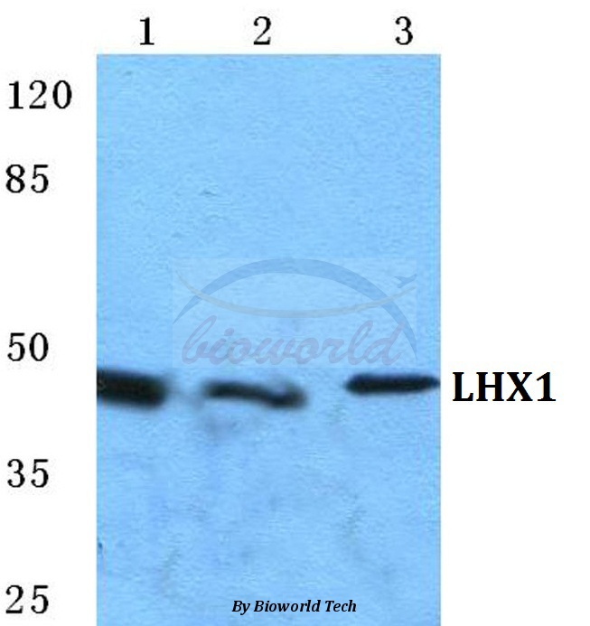
Figure 1. Western blot analysis of LHX1 using anti-LHX1 antibody (A05816-1). Electrophoresis was performed on a 5-20% SDS-PAGE gel at 70V (Stacking gel) / 90V (Resolving gel) for 2-3 hours. The sample well of each lane was loaded with 50ug of sample under reducing conditions. Lane 1: mouse brain tissue lysates. After Electrophoresis, proteins were transferred to a Nitrocellulose membrane at 150mA for 50-90 minutes. Blocked the membrane with 5% Non-fat Milk/ TBS for 1.5 hour at RT. The membrane was incubated with rabbit anti-LHX1 antigen affinity purified polyclonal antibody (Catalog # A05816-1) at 0.5 microg/mL overnight at 4°C, then washed with TBS-0.1%Tween 3 times with 5 minutes each and probed with a goat anti-rabbit IgG-HRP secondary antibody at a dilution of 1:5000 for 1.5 hour at RT. The signal is developed using an Enhanced Chemiluminescent detection (ECL) kit (Catalog # EK1002) with Tanon 5200 system. A specific band was detected for LHX1 at approximately 43KD. The expected band size for LHX1 is at 43KD.
. LHX1 was detected in paraffin-embedded section of mouse brain tissue. Heat mediated antigen retrieval was performed in EDTA buffer (pH8.0, epitope retrieval solution). The tissue section was blocked with 10% goat serum. The tissue section was then incubated with 1microg/ml rabbit anti-LHX1 Antibody (A05816-1) overnight at 4°C. Biotinylated goat anti-rabbit IgG was used as secondary antibody and incubated for 30 minutes at 37°C. The tissue section was developed using Strepavidin-Biotin-Complex (SABC) (Catalog # SA1022) with DAB as the chromogen.")
. Overlay histogram showing PC-3 cells stained with A05816-1 (Blue line). To facilitate intracellular staining, cells were fixed with 4% paraformaldehyde and permeabilized with permeabilization buffer. The cells were blocked with 10% normal goat serum. And then incubated with rabbit anti-LHX1 Antibody (A05816-1, 1microg/1x106 cells) for 30 min at 20°C. DyLight®488 conjugated goat anti-rabbit IgG (BA1127, 5-10microg/1x106 cells) was used as secondary antibody for 30 minutes at 20°C. Isotype control antibody (Green line) was rabbit IgG (1microg/1x106) used under the same conditions. Unlabelled sample without incubation with primary antibody and secondary antibody (Red line) was used as a blank control.")
Figure 1. Western blot analysis of LHX1 using anti-LHX1 antibody (A05816-1). Electrophoresis was performed on a 5-20% SDS-PAGE gel at 70V (Stacking gel) / 90V (Resolving gel) for 2-3 hours. The sample well of each lane was loaded with 50ug of sample under reducing conditions. Lane 1: mouse brain tissue lysates. After Electrophoresis, proteins were transferred to a Nitrocellulose membrane at 150mA for 50-90 minutes. Blocked the membrane with 5% Non-fat Milk/ TBS for 1.5 hour at RT. The membrane was incubated with rabbit anti-LHX1 antigen affinity purified polyclonal antibody (Catalog # A05816-1) at 0.5 microg/mL overnight at 4°C, then washed with TBS-0.1%Tween 3 times with 5 minutes each and probed with a goat anti-rabbit IgG-HRP secondary antibody at a dilution of 1:5000 for 1.5 hour at RT. The signal is developed using an Enhanced Chemiluminescent detection (ECL) kit (Catalog # EK1002) with Tanon 5200 system. A specific band was detected for LHX1 at approximately 43KD. The expected band size for LHX1 is at 43KD.
Anti-LIM1/LHX1 Antibody Picoband(r)
A05816-1-CARRIER-FREE
ApplicationsFlow Cytometry, Western Blot, ELISA, ImmunoHistoChemistry
Product group Antibodies
ReactivityHuman, Mouse
TargetLHX1
Overview
- SupplierBoster Bio
- Product NameAnti-LIM1/LHX1 Antibody Picoband(r)
- Delivery Days Customer9
- ApplicationsFlow Cytometry, Western Blot, ELISA, ImmunoHistoChemistry
- CertificationResearch Use Only
- ClonalityPolyclonal
- Concentration500 ug/ml
- Gene ID3975
- Target nameLHX1
- Target descriptionLIM homeobox 1
- Target synonymsLIM-1, LIM1, LIM/homeobox protein Lhx1, LIM homeobox protein 1, homeobox protein Lim-1
- HostRabbit
- IsotypeIgG
- Protein IDP48742
- Protein NameLIM/homeobox protein Lhx1
- Scientific DescriptionBoster Bio Anti-LIM1/LHX1 Antibody Picoband® catalog # A05816-1. Tested in ELISA, Flow Cytometry, IHC, WB applications. This antibody reacts with Human, Mouse. The brand Picoband indicates this is a premium antibody that guarantees superior quality, high affinity, and strong signals with minimal background in Western blot applications. Only our best-performing antibodies are designated as Picoband, ensuring unmatched performance.
- ReactivityHuman, Mouse
- Storage Instruction-20°C,2°C to 8°C
- UNSPSC12352203
Related products
Product group Antibodies
Anti-LHX1 AntibodyA28025
ApplicationsWestern Blot, ImmunoCytoChemistry, ImmunoHistoChemistry
ReactivityHuman, Mouse, Rat
- SizePrice
Product group Antibodies
Anti-LHX1 Antibody144-64893
ApplicationsWestern Blot
ReactivityHuman, Mouse, Rat
TargetLHX1
- SizePrice
Product group Antibodies
LHX1 AntibodyCSB-PA009825
ApplicationsWestern Blot, ELISA, ImmunoHistoChemistry
ReactivityHuman, Mouse, Rat
TargetLHX1
- SizePrice
Product group Antibodies

LHX1 Antibody (Internal)LS-C384339
ApplicationsWestern Blot, ELISA, ImmunoHistoChemistry
ReactivityHuman, Mouse, Rat
TargetLHX1
- SizePrice

Product group Antibodies
Anti-LHX1 AntibodyHPA073521
ApplicationsImmunoHistoChemistry
ReactivityHuman
TargetLHX1
- SizePrice
![LIM1 antibody detects LIM1 protein at cytoplasm and nucleus by immunofluorescent analysis. Sample: DIV9 rat E18 primary cortical neuron cells were fixed in 4% paraformaldehyde at RT for 15 min. Green: LIM1 stained by LIM1 antibody (GTX129215) diluted at 1:250. Red: Tau, an axon marker, stained by Tau antibody [GT287] (GTX634809) diluted at 1:500. Blue: Fluoroshield with DAPI (GTX30920).](https://www.genetex.com/upload/website/prouct_img/normal/GTX129215/GTX129215_44748_20231222_ICC_IF_R_24011618_418.webp)
Product group Antibodies
LIM1 antibodyGTX129215
ApplicationsImmunoFluorescence, ImmunoCytoChemistry
ReactivityHuman, Rat
TargetLHX1
- SizePrice