


Figure 1. Western blot analysis of LIS1/PAFAH1B1 using anti-LIS1/PAFAH1B1 antibody (A01273-1). Electrophoresis was performed on a 5-20% SDS-PAGE gel at 70V (Stacking gel) / 90V (Resolving gel) for 2-3 hours. The sample well of each lane was loaded with 30 ug of sample under reducing conditions. Lane 1: human U87 whole cell lysates. After electrophoresis, proteins were transferred to a nitrocellulose membrane at 150 mA for 50-90 minutes. Blocked the membrane with 5% non-fat milk/TBS for 1.5 hour at RT. The membrane was incubated with rabbit anti-LIS1/PAFAH1B1 antigen affinity purified polyclonal antibody (Catalog # A01273-1) at 0.5 microg/mL overnight at 4°C, then washed with TBS-0.1%Tween 3 times with 5 minutes each and probed with a goat anti-rabbit IgG-HRP secondary antibody at a dilution of 1:5000 for 1.5 hour at RT. The signal is developed using an Enhanced Chemiluminescent detection (ECL) kit (Catalog # EK1002) with Tanon 5200 system. A specific band was detected for LIS1/PAFAH1B1 at approximately 47 kDa. The expected band size for LIS1/PAFAH1B1 is at 47 kDa.
Figure 1. Western blot analysis of LIS1/PAFAH1B1 using anti-LIS1/PAFAH1B1 antibody (A01273-1). Electrophoresis was performed on a 5-20% SDS-PAGE gel at 70V (Stacking gel) / 90V (Resolving gel) for 2-3 hours. The sample well of each lane was loaded with 30 ug of sample under reducing conditions. Lane 1: human U87 whole cell lysates. After electrophoresis, proteins were transferred to a nitrocellulose membrane at 150 mA for 50-90 minutes. Blocked the membrane with 5% non-fat milk/TBS for 1.5 hour at RT. The membrane was incubated with rabbit anti-LIS1/PAFAH1B1 antigen affinity purified polyclonal antibody (Catalog # A01273-1) at 0.5 microg/mL overnight at 4°C, then washed with TBS-0.1%Tween 3 times with 5 minutes each and probed with a goat anti-rabbit IgG-HRP secondary antibody at a dilution of 1:5000 for 1.5 hour at RT. The signal is developed using an Enhanced Chemiluminescent detection (ECL) kit (Catalog # EK1002) with Tanon 5200 system. A specific band was detected for LIS1/PAFAH1B1 at approximately 47 kDa. The expected band size for LIS1/PAFAH1B1 is at 47 kDa.
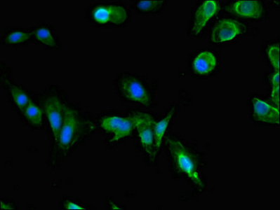
Anti-LIS1/PAFAH1B1 Antibody Picoband(r)
A01273-1-DYLIGHT488
ApplicationsWestern Blot, ELISA
Product group Antibodies
ReactivityHuman
TargetPAFAH1B1
Overview
- SupplierBoster Bio
- Product NameAnti-LIS1/PAFAH1B1 Antibody Picoband(r)
- Delivery Days Customer9
- ApplicationsWestern Blot, ELISA
- CertificationResearch Use Only
- ClonalityPolyclonal
- Concentration500 ug/ml
- ConjugateDyLight 488
- Gene ID5048
- Target namePAFAH1B1
- Target descriptionplatelet activating factor acetylhydrolase 1b regulatory subunit 1
- Target synonymsLIS1, LIS2, MDCR, MDS, NudF, PAFAH, platelet-activating factor acetylhydrolase IB subunit beta, lissencephaly 1 protein, platelet-activating factor acetylhydrolase 1b, regulatory subunit 1 (45kDa)
- HostRabbit
- IsotypeIgG
- Protein IDP43034
- Protein NamePlatelet-activating factor acetylhydrolase IB subunit beta
- Scientific DescriptionBoster Bio Anti-LIS1/PAFAH1B1 Antibody Picoband® catalog # A01273-1. Tested in ELISA, WB applications. This antibody reacts with Human. The brand Picoband indicates this is a premium antibody that guarantees superior quality, high affinity, and strong signals with minimal background in Western blot applications. Only our best-performing antibodies are designated as Picoband, ensuring unmatched performance.
- ReactivityHuman
- Storage Instruction-20°C,2°C to 8°C
- UNSPSC12352203
Related products
Product group Antibodies
PAFAH1B1 AntibodyCSB-PA017383LA01HU
ApplicationsImmunoFluorescence, ELISA
ReactivityHuman
TargetPAFAH1B1
- SizePrice
Product group Antibodies
Anti-LIS1/PAFAH1B1 Antibody Picoband(r)A01273-1-CARRIER-FREE
ApplicationsWestern Blot, ELISA
ReactivityHuman
TargetPAFAH1B1
- SizePrice
Product group Antibodies

LIS1 AntibodyABX430259
ApplicationsWestern Blot, ELISA, ImmunoHistoChemistry
- SizePrice
Product group Antibodies
Anti-PAFAH1B1 AntibodyA45000
ApplicationsImmunoHistoChemistry
ReactivityHuman, Mouse, Rat
- SizePrice
Product group Antibodies

PAFAH1B1 / LIS1 AntibodyLS-C747734
ApplicationsWestern Blot
ReactivityHuman, Mouse
TargetPAFAH1B1
- SizePrice
Product group Antibodies

Goat anti-LIS1 / PAFAH1B1EB05439
ApplicationsWestern Blot, ELISA, ImmunoHistoChemistry
ReactivityHuman, Rat
TargetPAFAH1B1
- SizePrice

Product group Antibodies
Anti-PAFAH1B1 AntibodyHPA020036
ApplicationsImmunoCytoChemistry, ImmunoHistoChemistry
ReactivityHuman
TargetPAFAH1B1
- SizePrice
Product group Antibodies
ApplicationsFlow Cytometry, Western Blot, ImmunoCytoChemistry
ReactivityHuman, Mouse, Rat
TargetPAFAH1B1
- SizePrice
Product group Antibodies
LIS1 antibodyGTX16993
ApplicationsImmunoFluorescence, Western Blot, ELISA, ImmunoCytoChemistry
ReactivityHuman, Mouse, Rat
TargetPAFAH1B1
- SizePrice