

Anti-LMO7 Antibody
A28953
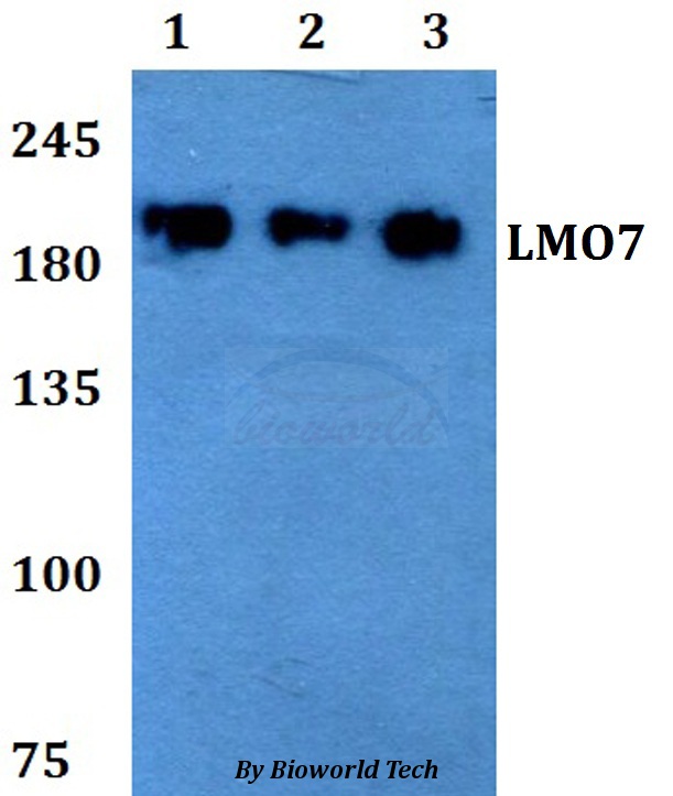
ApplicationsWestern Blot
Product group Antibodies
ReactivityHuman, Mouse, Rat
Overview
- SupplierAntibodies.com
- Product NameAnti-LMO7 Antibody
- Delivery Days Customer7
- ApplicationsWestern Blot
- CertificationResearch Use Only
- ClonalityPolyclonal
- ConjugateUnconjugated
- Estimated Purity>95%
- HostRabbit
- Scientific DescriptionRabbit polyclonal antibody to LMO7
- ReactivityHuman, Mouse, Rat
- UNSPSC12352203
Related products

Product group Antibodies
Anti-LMO7 Antibody Picoband(r)A05712-2-CARRIER-FREE
ApplicationsFlow Cytometry, Western Blot, ELISA
ReactivityHuman
TargetLMO7
- SizePrice
Product group Antibodies

Goat anti-LMO7 / FBXO20EB06212
ApplicationsWestern Blot, ELISA
ReactivityCanine, Human, Mouse, Rat
TargetLMO7
- SizePrice

Product group Antibodies
Anti-LMO7 AntibodyHPA050184
ApplicationsImmunoCytoChemistry
ReactivityHuman
TargetLMO7
- SizePrice
Product group Antibodies

LMO7 AntibodyLS-C680776
ApplicationsImmunoFluorescence, ELISA, ImmunoHistoChemistry, ImmunoHistoChemistry Paraffin
ReactivityHuman
TargetLMO7
- SizePrice
Product group Antibodies
LMO7 AntibodyCSB-PA823897LA01HU
ApplicationsImmunoFluorescence, ELISA, ImmunoHistoChemistry
ReactivityHuman
TargetLMO7
- SizePrice
Product group Antibodies
LMO7 antibody, InternalGTX25955
ApplicationsWestern Blot
ReactivityHuman
TargetLMO7
- SizePrice
Product group Antibodies
LMO7 AntibodyPACO60236
ApplicationsImmunoFluorescence, ELISA, ImmunoHistoChemistry
ReactivityHuman
TargetLMO7
- SizePrice