


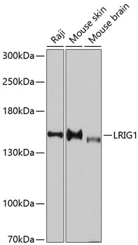
Figure 1. Western blot analysis of LRIG1 using anti-LRIG1 antibody (A04613-2). Electrophoresis was performed on a 5-20% SDS-PAGE gel at 70V (Stacking gel) / 90V (Resolving gel) for 2-3 hours. The sample well of each lane was loaded with 30 ug of sample under reducing conditions. Lane 1: human 293T whole cell lysates. After electrophoresis, proteins were transferred to a nitrocellulose membrane at 150 mA for 50-90 minutes. Blocked the membrane with 5% non-fat milk/TBS for 1.5 hour at RT. The membrane was incubated with rabbit anti-LRIG1 antigen affinity purified polyclonal antibody (Catalog # A04613-2) at 0.5 microg/mL overnight at 4°C, then washed with TBS-0.1%Tween 3 times with 5 minutes each and probed with a goat anti-rabbit IgG-HRP secondary antibody at a dilution of 1:5000 for 1.5 hour at RT. The signal is developed using an Enhanced Chemiluminescent detection (ECL) kit (Catalog # EK1002) with Tanon 5200 system. A specific band was detected for LRIG1 at approximately 145 kDa. The expected band size for LRIG1 is at 119 kDa.
Figure 1. Western blot analysis of LRIG1 using anti-LRIG1 antibody (A04613-2). Electrophoresis was performed on a 5-20% SDS-PAGE gel at 70V (Stacking gel) / 90V (Resolving gel) for 2-3 hours. The sample well of each lane was loaded with 30 ug of sample under reducing conditions. Lane 1: human 293T whole cell lysates. After electrophoresis, proteins were transferred to a nitrocellulose membrane at 150 mA for 50-90 minutes. Blocked the membrane with 5% non-fat milk/TBS for 1.5 hour at RT. The membrane was incubated with rabbit anti-LRIG1 antigen affinity purified polyclonal antibody (Catalog # A04613-2) at 0.5 microg/mL overnight at 4°C, then washed with TBS-0.1%Tween 3 times with 5 minutes each and probed with a goat anti-rabbit IgG-HRP secondary antibody at a dilution of 1:5000 for 1.5 hour at RT. The signal is developed using an Enhanced Chemiluminescent detection (ECL) kit (Catalog # EK1002) with Tanon 5200 system. A specific band was detected for LRIG1 at approximately 145 kDa. The expected band size for LRIG1 is at 119 kDa.
Anti-LRIG1 Antibody Picoband(r)
A04613-2-CARRIER-FREE
ApplicationsWestern Blot, ELISA
Product group Antibodies
ReactivityHuman
TargetLRIG1
Overview
- SupplierBoster Bio
- Product NameAnti-LRIG1 Antibody Picoband(r)
- Delivery Days Customer9
- ApplicationsWestern Blot, ELISA
- CertificationResearch Use Only
- ClonalityPolyclonal
- Concentration500 ug/ml
- Gene ID26018
- Target nameLRIG1
- Target descriptionleucine rich repeats and immunoglobulin like domains 1
- Target synonymsLIG-1, LIG1, leucine-rich repeats and immunoglobulin-like domains protein 1, leucine-rich repeat protein LRIG1, ortholog of mouse integral membrane glycoprotein LIG-1
- HostRabbit
- Protein IDQ96JA1
- Protein NameLeucine-rich repeats and immunoglobulin-like domains protein 1
- Scientific DescriptionBoster Bio Anti-LRIG1 Antibody Picoband® catalog # A04613-2. Tested in WB, ELISA applications. This antibody reacts with Human. The brand Picoband indicates this is a premium antibody that guarantees superior quality, high affinity, and strong signals with minimal background in Western blot applications. Only our best-performing antibodies are designated as Picoband, ensuring unmatched performance.
- ReactivityHuman
- Storage Instruction-20°C,2°C to 8°C
- UNSPSC12352203
Related products
Product group Antibodies
Anti-LRIG1 AntibodyA11278
ApplicationsWestern Blot
ReactivityHuman, Mouse
- SizePrice
Product group Antibodies
Anti-LRIG1 Antibody144-10297
ApplicationsWestern Blot
ReactivityHuman, Mouse
TargetLRIG1
- SizePrice

Product group Antibodies
References
LRIG1 Polyclonal AntibodyBS-1844R
ApplicationsImmunoFluorescence, Western Blot, ELISA, ImmunoCytoChemistry, ImmunoHistoChemistry, ImmunoHistoChemistry Frozen, ImmunoHistoChemistry Paraffin
ReactivityHuman, Mouse, Rat
TargetLRIG1
- SizePrice
Product group Antibodies
ApplicationsImmunoPrecipitation, Western Blot, ImmunoCytoChemistry, ImmunoHistoChemistry
ReactivityPorcine
TargetLRIG1
- SizePrice
Product group Antibodies
LRIG1 AntibodyCSB-PA245012
ApplicationsELISA, ImmunoHistoChemistry
ReactivityHuman
TargetLRIG1
- SizePrice
Product group Antibodies

LRIG1 AntibodyLS-C402418
ApplicationsELISA, ImmunoHistoChemistry
ReactivityHuman
TargetLRIG1
- SizePrice

Product group Antibodies
Anti-LRIG1 AntibodyHPA011846
ApplicationsWestern Blot, ImmunoCytoChemistry, ImmunoHistoChemistry
ReactivityHuman
TargetLRIG1
- SizePrice
Product group Antibodies
LRIG1 antibody [C2C3], C-termGTX119485
ApplicationsImmunoPrecipitation, Western Blot, ImmunoHistoChemistry, ImmunoHistoChemistry Paraffin
ReactivityHuman, Mouse
TargetLRIG1
- SizePrice
Product group Antibodies
Anti-LRIG1 AntibodyCAB10297
ApplicationsWestern Blot, ELISA
ReactivityHuman
TargetLRIG1
- SizePrice