

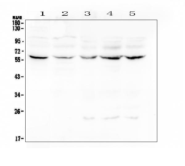
Figure 1. Western blot analysis of Lumican using anti-Lumican antibody (A01034-1). Electrophoresis was performed on a 5-20% SDS-PAGE gel at 70V (Stacking gel) / 90V (Resolving gel) for 2-3 hours. The sample well of each lane was loaded with 50ug of sample under reducing conditions. Lane 1: human placenta tissue lysates, Lane 2: human Caco-2 whole cell lysate, Lane 3: human CCRF-CEM whole cell lysate, Lane 4: human Hela whole cell lysate, Lane 5: human Jurkat whole cell lysate. After Electrophoresis, proteins were transferred to a Nitrocellulose membrane at 150mA for 50-90 minutes. Blocked the membrane with 5% Non-fat Milk/ TBS for 1.5 hour at RT. The membrane was incubated with rabbit anti-Lumican antigen affinity purified polyclonal antibody (Catalog # A01034-1) at 0.5 microg/mL overnight at 4°C, then washed with TBS-0.1%Tween 3 times with 5 minutes each and probed with a goat anti-rabbit IgG-HRP secondary antibody at a dilution of 1:10000 for 1.5 hour at RT. The signal is developed using an Enhanced Chemiluminescent detection (ECL) kit (Catalog # EK1002) with Tanon 5200 system. A specific band was detected for Lumican at approximately 58KD. The expected band size for Lumican is at 38KD.
. Electrophoresis was performed on a 5-20% SDS-PAGE gel at 70V (Stacking gel) / 90V (Resolving gel) for 2-3 hours. The sample well of each lane was loaded with 50ug of sample under reducing conditions. Lane 1: mouse liver tissue lysates, Lane 2: mouse ovary tissue lysates, Lane 3: mouse testis tissue lysates, Lane 4: mouse lung tissue lysates, Lane 5: rat liver tissue lysates, Lane 6: rat ovary tissue lysates, Lane 7: rat testis tissue lysates, Lane 8: rat lung tissue lysates, Lane 9: rat heart tissue lysates. After Electrophoresis, proteins were transferred to a Nitrocellulose membrane at 150mA for 50-90 minutes. Blocked the membrane with 5% Non-fat Milk/ TBS for 1.5 hour at RT. The membrane was incubated with rabbit anti-Lumican antigen affinity purified polyclonal antibody (Catalog # A01034-1) at 0.5 microg/mL overnight at 4°C, then washed with TBS-0.1%Tween 3 times with 5 minutes each and probed with a goat anti-rabbit IgG-HRP secondary antibody at a dilution of 1:10000 for 1.5 hour at RT. The signal is developed using an Enhanced Chemiluminescent detection (ECL) kit (Catalog # EK1002) with Tanon 5200 system. A specific band was detected for Lumican at approximately 58KD. The expected band size for Lumican is at 38KD.")
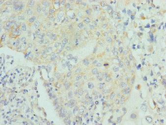
. Lumican was detected in paraffin-embedded section of human ovary cancer tissue. Heat mediated antigen retrieval was performed in citrate buffer (pH6, epitope retrieval solution) for 20 mins. The tissue section was blocked with 10% goat serum. The tissue section was then incubated with 1microg/ml rabbit anti-Lumican Antibody (A01034-1) overnight at 4°C. Biotinylated goat anti-rabbit IgG was used as secondary antibody and incubated for 30 minutes at 37°C. The tissue section was developed using Strepavidin-Biotin-Complex (SABC)(Catalog # SA1022) with DAB as the chromogen.")
. Lumican was detected in paraffin-embedded section of human pancreatic cancer tissue. Heat mediated antigen retrieval was performed in citrate buffer (pH6, epitope retrieval solution) for 20 mins. The tissue section was blocked with 10% goat serum. The tissue section was then incubated with 1microg/ml rabbit anti-Lumican Antibody (A01034-1) overnight at 4°C. Biotinylated goat anti-rabbit IgG was used as secondary antibody and incubated for 30 minutes at 37°C. The tissue section was developed using Strepavidin-Biotin-Complex (SABC)(Catalog # SA1022) with DAB as the chromogen.")
. Lumican was detected in paraffin-embedded section of human lung cancer tissue. Heat mediated antigen retrieval was performed in citrate buffer (pH6, epitope retrieval solution) for 20 mins. The tissue section was blocked with 10% goat serum. The tissue section was then incubated with 1microg/ml rabbit anti-Lumican Antibody (A01034-1) overnight at 4°C. Biotinylated goat anti-rabbit IgG was used as secondary antibody and incubated for 30 minutes at 37°C. The tissue section was developed using Strepavidin-Biotin-Complex (SABC)(Catalog # SA1022) with DAB as the chromogen.")
. Lumican was detected in paraffin-embedded section of human placenta tissue. Heat mediated antigen retrieval was performed in citrate buffer (pH6, epitope retrieval solution) for 20 mins. The tissue section was blocked with 10% goat serum. The tissue section was then incubated with 1microg/ml rabbit anti-Lumican Antibody (A01034-1) overnight at 4°C. Biotinylated goat anti-rabbit IgG was used as secondary antibody and incubated for 30 minutes at 37°C. The tissue section was developed using Strepavidin-Biotin-Complex (SABC)(Catalog # SA1022) with DAB as the chromogen.")
Figure 1. Western blot analysis of Lumican using anti-Lumican antibody (A01034-1). Electrophoresis was performed on a 5-20% SDS-PAGE gel at 70V (Stacking gel) / 90V (Resolving gel) for 2-3 hours. The sample well of each lane was loaded with 50ug of sample under reducing conditions. Lane 1: human placenta tissue lysates, Lane 2: human Caco-2 whole cell lysate, Lane 3: human CCRF-CEM whole cell lysate, Lane 4: human Hela whole cell lysate, Lane 5: human Jurkat whole cell lysate. After Electrophoresis, proteins were transferred to a Nitrocellulose membrane at 150mA for 50-90 minutes. Blocked the membrane with 5% Non-fat Milk/ TBS for 1.5 hour at RT. The membrane was incubated with rabbit anti-Lumican antigen affinity purified polyclonal antibody (Catalog # A01034-1) at 0.5 microg/mL overnight at 4°C, then washed with TBS-0.1%Tween 3 times with 5 minutes each and probed with a goat anti-rabbit IgG-HRP secondary antibody at a dilution of 1:10000 for 1.5 hour at RT. The signal is developed using an Enhanced Chemiluminescent detection (ECL) kit (Catalog # EK1002) with Tanon 5200 system. A specific band was detected for Lumican at approximately 58KD. The expected band size for Lumican is at 38KD.
Anti-Lumican Antibody Picoband(r)
A01034-1-CARRIER-FREE
ApplicationsWestern Blot, ELISA, ImmunoHistoChemistry
Product group Antibodies
ReactivityHuman, Mouse, Rat
TargetLUM
Overview
- SupplierBoster Bio
- Product NameAnti-Lumican Antibody Picoband(r)
- Delivery Days Customer9
- ApplicationsWestern Blot, ELISA, ImmunoHistoChemistry
- CertificationResearch Use Only
- ClonalityPolyclonal
- Concentration500 ug/ml
- Gene ID4060
- Target nameLUM
- Target descriptionlumican
- Target synonymsLDC, SLRR2D, lumican, KSPG lumican, epididymis secretory sperm binding protein, keratan sulfate proteoglycan lumican, lumican proteoglycan
- HostRabbit
- IsotypeIgG
- Protein IDP51884
- Protein NameLumican
- Scientific DescriptionBoster Bio Anti-Lumican Antibody Picoband® catalog # A01034-1. Tested in ELISA, IHC, WB applications. This antibody reacts with Human, Mouse, Rat. The brand Picoband indicates this is a premium antibody that guarantees superior quality, high affinity, and strong signals with minimal background in Western blot applications. Only our best-performing antibodies are designated as Picoband, ensuring unmatched performance.
- ReactivityHuman, Mouse, Rat
- Storage Instruction-20°C,2°C to 8°C
- UNSPSC12352203
Related products
Product group Antibodies
Anti-LUM AntibodyA30749
ApplicationsWestern Blot, ImmunoHistoChemistry
ReactivityHuman, Mouse, Rat
- SizePrice
Product group Antibodies
Anti-LUM Antibody144-05352
ApplicationsWestern Blot
ReactivityHuman, Mouse
TargetLUM
- SizePrice
Product group Antibodies

Lumican (LUM) AntibodyABX213779
ApplicationsELISA, ImmunoHistoChemistry
- SizePrice
Product group Antibodies
References
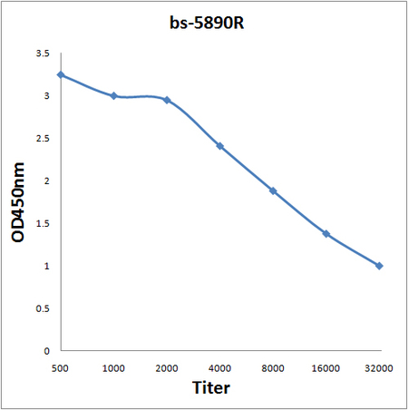
Lumican Polyclonal AntibodyBS-5890R
ApplicationsImmunoFluorescence, Western Blot, ELISA, ImmunoCytoChemistry, ImmunoHistoChemistry, ImmunoHistoChemistry Frozen, ImmunoHistoChemistry Paraffin
ReactivityBovine, Equine, Human, Mouse, Porcine, Rabbit, Rat
TargetLUM
- SizePrice
Product group Antibodies
LUM AntibodyCSB-PA013234ESR1HU
ApplicationsELISA, ImmunoHistoChemistry
ReactivityHuman
TargetLUM
- SizePrice
Product group Antibodies
ApplicationsImmunoPrecipitation, Western Blot, ImmunoCytoChemistry, ImmunoHistoChemistry
TargetLUM
- SizePrice
Product group Antibodies

Lumican AntibodyLS-C488192
ApplicationsFlow Cytometry, ImmunoFluorescence, ImmunoCytoChemistry
ReactivityHuman
TargetLUM
- SizePrice

Product group Antibodies
Anti-LUM AntibodyHPA001522
ApplicationsImmunoHistoChemistry
ReactivityHuman
TargetLUM
- SizePrice
Product group Antibodies
LUM antibodyGTX131377
ApplicationsWestern Blot, ImmunoHistoChemistry, ImmunoHistoChemistry Paraffin
ReactivityHuman, Mouse
TargetLUM
- SizePrice
Product group Antibodies
ApplicationsWestern Blot, ELISA, ImmunoHistoChemistry, ImmunoHistoChemistry Paraffin
ReactivityHuman
TargetLUM
- SizePrice