


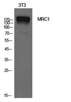
Figure 1. Western blot analysis of MRC1 using anti-MRC1 antibody (A02285-2). Electrophoresis was performed on a 5-20% SDS-PAGE gel at 70V (Stacking gel) / 90V (Resolving gel) for 2-3 hours. The sample well of each lane was loaded with 30 ug of sample under reducing conditions. Lane 1: rat lung tissue lysates, Lane 2: mouse lung tissue lysates, Lane 3: human liver tissue lysates, Lane 4: monkey lung tissue lysates. After electrophoresis, proteins were transferred to a nitrocellulose membrane at 150 mA for 50-90 minutes. Blocked the membrane with 5% non-fat milk/TBS for 1.5 hour at RT. The membrane was incubated with rabbit anti-MRC1 antigen affinity purified polyclonal antibody (Catalog # A02285-2) at 0.5 microg/mL overnight at 4°C, then washed with TBS-0.1%Tween 3 times with 5 minutes each and probed with a goat anti-rabbit IgG-HRP secondary antibody at a dilution of 1:5000 for 1.5 hour at RT. The signal is developed using an Enhanced Chemiluminescent detection (ECL) kit (Catalog # EK1002) with Tanon 5200 system. A specific band was detected for MRC1 at approximately 190-200 kDa. The expected band size for MRC1 is at 166 kDa.
. MRC1 was detected in a paraffin-embedded section of human liver cancer tissue. Heat mediated antigen retrieval was performed in EDTA buffer (pH 8.0, epitope retrieval solution). The tissue section was blocked with 10% goat serum. The tissue section was then incubated with 2 microg/ml rabbit anti-MRC1 Antibody (A02285-2) overnight at 4°C. Peroxidase Conjugated Goat Anti-rabbit IgG was used as secondary antibody and incubated for 30 minutes at 37°C. The tissue section was developed using HRP Conjugated Rabbit IgG Super Vision Assay Kit (Catalog # SV0002) with DAB as the chromogen.")
. MRC1 was detected in a paraffin-embedded section of human tonsil tissue. Heat mediated antigen retrieval was performed in EDTA buffer (pH 8.0, epitope retrieval solution). The tissue section was blocked with 10% goat serum. The tissue section was then incubated with 2 microg/ml rabbit anti-MRC1 Antibody (A02285-2) overnight at 4°C. Peroxidase Conjugated Goat Anti-rabbit IgG was used as secondary antibody and incubated for 30 minutes at 37°C. The tissue section was developed using HRP Conjugated Rabbit IgG Super Vision Assay Kit (Catalog # SV0002) with DAB as the chromogen.")
. MRC1 was detected in a paraffin-embedded section of mouse liver tissue. Heat mediated antigen retrieval was performed in EDTA buffer (pH 8.0, epitope retrieval solution). The tissue section was blocked with 10% goat serum. The tissue section was then incubated with 2 microg/ml rabbit anti-MRC1 Antibody (A02285-2) overnight at 4°C. Peroxidase Conjugated Goat Anti-rabbit IgG was used as secondary antibody and incubated for 30 minutes at 37°C. The tissue section was developed using HRP Conjugated Rabbit IgG Super Vision Assay Kit (Catalog # SV0002) with DAB as the chromogen.")
. MRC1 was detected in a paraffin-embedded section of rat liver tissue. Heat mediated antigen retrieval was performed in EDTA buffer (pH 8.0, epitope retrieval solution). The tissue section was blocked with 10% goat serum. The tissue section was then incubated with 2 microg/ml rabbit anti-MRC1 Antibody (A02285-2) overnight at 4°C. Peroxidase Conjugated Goat Anti-rabbit IgG was used as secondary antibody and incubated for 30 minutes at 37°C. The tissue section was developed using HRP Conjugated Rabbit IgG Super Vision Assay Kit (Catalog # SV0002) with DAB as the chromogen.")
. MRC1 was detected in a paraffin-embedded section of human tonsil tissue. Heat mediated antigen retrieval was performed in EDTA buffer (pH 8.0, epitope retrieval solution). The tissue section was blocked with 10% goat serum. The tissue section was then incubated with 5 microg/mL rabbit anti-MRC1 Antibody (A02285-2) overnight at 4°C. DyLight®550 Conjugated Goat Anti-Rabbit IgG (BA1135) was used as secondary antibody at 1:500 dilution and incubated for 30 minutes at 37°C. The section was counterstained with DAPI. Visualize using a fluorescence microscope and filter sets appropriate for the label used.")
. MRC1 was detected in a paraffin-embedded section of mouse liver tissue. Heat mediated antigen retrieval was performed in EDTA buffer (pH 8.0, epitope retrieval solution). The tissue section was blocked with 10% goat serum. The tissue section was then incubated with 5 microg/mL rabbit anti-MRC1 Antibody (A02285-2) overnight at 4°C. DyLight®550 Conjugated Goat Anti-Rabbit IgG (BA1135) was used as secondary antibody at 1:500 dilution and incubated for 30 minutes at 37°C. The section was counterstained with DAPI. Visualize using a fluorescence microscope and filter sets appropriate for the label used.")
Figure 1. Western blot analysis of MRC1 using anti-MRC1 antibody (A02285-2). Electrophoresis was performed on a 5-20% SDS-PAGE gel at 70V (Stacking gel) / 90V (Resolving gel) for 2-3 hours. The sample well of each lane was loaded with 30 ug of sample under reducing conditions. Lane 1: rat lung tissue lysates, Lane 2: mouse lung tissue lysates, Lane 3: human liver tissue lysates, Lane 4: monkey lung tissue lysates. After electrophoresis, proteins were transferred to a nitrocellulose membrane at 150 mA for 50-90 minutes. Blocked the membrane with 5% non-fat milk/TBS for 1.5 hour at RT. The membrane was incubated with rabbit anti-MRC1 antigen affinity purified polyclonal antibody (Catalog # A02285-2) at 0.5 microg/mL overnight at 4°C, then washed with TBS-0.1%Tween 3 times with 5 minutes each and probed with a goat anti-rabbit IgG-HRP secondary antibody at a dilution of 1:5000 for 1.5 hour at RT. The signal is developed using an Enhanced Chemiluminescent detection (ECL) kit (Catalog # EK1002) with Tanon 5200 system. A specific band was detected for MRC1 at approximately 190-200 kDa. The expected band size for MRC1 is at 166 kDa.
Anti-Mannose Receptor/MRC1 Picoband(r) Antibody
A02285-2-CARRIER-FREE
ApplicationsImmunoFluorescence, Western Blot, ELISA, ImmunoHistoChemistry
Product group Antibodies
ReactivityHuman, Monkey, Mouse, Rat
TargetMRC1
Overview
- SupplierBoster Bio
- Product NameAnti-Mannose Receptor/MRC1 Picoband(r) Antibody
- Delivery Days Customer9
- ApplicationsImmunoFluorescence, Western Blot, ELISA, ImmunoHistoChemistry
- CertificationResearch Use Only
- ClonalityPolyclonal
- Concentration500 ug/ml
- Gene ID4360
- Target nameMRC1
- Target descriptionmannose receptor C-type 1
- Target synonymsCD206, CLEC13D, CLEC13DL, MMR, MRC1L1, bA541I19.1, hMR, macrophage mannose receptor 1, C-type lectin domain family 13 member D, human mannose receptor, macrophage mannose receptor 1-like protein 1, mannose receptor, C type 1-like 1
- HostRabbit
- IsotypeIgG
- Protein IDP22897
- Protein NameMacrophage mannose receptor 1
- Scientific DescriptionBoster Bio Anti-Mannose Receptor/MRC1 Picoband® Antibody catalog # A02285-2. Tested in ELISA, IF, IHC, WB applications. This antibody reacts with Human, Monkey, Mouse, Rat. The brand Picoband indicates this is a premium antibody that guarantees superior quality, high affinity, and strong signals with minimal background in Western blot applications. Only our best-performing antibodies are designated as Picoband, ensuring unmatched performance.
- ReactivityHuman, Monkey, Mouse, Rat
- Storage Instruction-20°C,2°C to 8°C
- UNSPSC12352203
Related products
Product group Antibodies

Anti-CD206 [6D5]Ab03382-1.1
ApplicationsELISA
ReactivityHuman
TargetMRC1
- SizePrice
Product group Antibodies
Anti-MRC1 AntibodyA101372
ApplicationsWestern Blot, ELISA
ReactivityHuman
- SizePrice
Product group Antibodies
Anti-MRC1 Antibody144-66582
ApplicationsWestern Blot, ImmunoHistoChemistry
ReactivityHuman, Mouse, Rat
TargetMRC1
- SizePrice


Product group Antibodies
Anti-MRC1 AntibodyAMAB90746
ApplicationsWestern Blot, ImmunoHistoChemistry
ReactivityHuman
TargetMRC1
- SizePrice
Product group Antibodies
MRC1 AntibodyCSB-PA006303
ApplicationsWestern Blot, ELISA
ReactivityHuman
TargetMRC1
- SizePrice
Product group Antibodies
ApplicationsImmunoPrecipitation, Western Blot, ImmunoCytoChemistry, ImmunoHistoChemistry
ReactivityMouse
TargetMRC1
- SizePrice
Product group Antibodies
References
MRC1 Polyclonal AntibodyBS-4727R
ApplicationsFlow Cytometry, Western Blot, ELISA
ReactivityHuman
TargetMRC1
- SizePrice
Product group Antibodies

ApplicationsFlow Cytometry, Western Blot, ImmunoHistoChemistry, ImmunoHistoChemistry Frozen
ReactivityHuman
TargetMRC1
- SizePrice
![IHC-Fr analysis of human tonsil tissue using GTX42266 Mannose Receptor antibody [7-450].](https://www.genetex.com/upload/website/prouct_img/normal/GTX42266/GTX42266_4774_IHC-Fr_w_23060820_199.webp)
Product group Antibodies
References
ApplicationsImmunoHistoChemistry, ImmunoHistoChemistry Frozen
ReactivityHuman
TargetMRC1
- SizePrice