

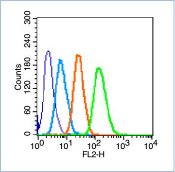
HepG2 cells(black) were incubated in 5% BSA blocking buffer for 30 min at room temperature. Cells were then stained with MRC1 Antibody(bs-4727R) at 1:50 dilution in blocking buffer and incubated for 30 min at room temperature, washed twice with 2% BSA in PBS. Acquisitions of 20,000 events were performed. Cells stained with primary antibody (green) and isotype control (orange).
at 1:50 dilution in blocking buffer and incubated for 30 min at room temperature, washed twice with 2%BSA in PBS, followed by secondary antibody incubation for 40 min at room temperature. Acquisitions of 20,000 events were performed. Cells stained with primary antibody (green), and isotype control (orange).")
HepG2 cells(black) were incubated in 5% BSA blocking buffer for 30 min at room temperature. Cells were then stained with MRC1 Antibody(bs-4727R) at 1:50 dilution in blocking buffer and incubated for 30 min at room temperature, washed twice with 2% BSA in PBS. Acquisitions of 20,000 events were performed. Cells stained with primary antibody (green) and isotype control (orange).
MRC1 Polyclonal Antibody
BS-4727R
ApplicationsFlow Cytometry, Western Blot, ELISA
Product group Antibodies
ReactivityHuman
TargetMRC1
Overview
- SupplierBioss
- Product NameMRC1 Polyclonal Antibody
- Delivery Days Customer16
- ApplicationsFlow Cytometry, Western Blot, ELISA
- Applications SupplierWB(1:300-5000), ELISA(1:500-1000), FCM(1:20-100)
- CertificationResearch Use Only
- ClonalityPolyclonal
- Concentration1 ug/ul
- ConjugateUnconjugated
- Gene ID4360
- Target nameMRC1
- Target descriptionmannose receptor C-type 1
- Target synonymsCD206, CLEC13D, CLEC13DL, MMR, MRC1L1, bA541I19.1, hMR, macrophage mannose receptor 1, C-type lectin domain family 13 member D, human mannose receptor, macrophage mannose receptor 1-like protein 1, mannose receptor, C type 1-like 1
- HostRabbit
- IsotypeIgG
- Protein IDP22897
- Protein NameMacrophage mannose receptor 1
- ReactivityHuman
- Storage Instruction-20°C
- UNSPSC41116161
References
- Sonodynamic therapy in atherosclerosis by curcumin nanosuspensions: Preparation design, efficacy evaluation, and mechanisms analysis. Jiang L et al., 2020 Jan, Eur J Pharm BiopharmRead this paper
- Structural characterization and immunomodulatory activity of a novel acid polysaccharide isolated from the pulp of Rosa laevigata Michx fruit. Zhan Q et al., 2020 Feb 15, Int J Biol MacromolRead this paper
- A Multi-Functional Silicon Nanoparticle Designed for Enhanced Osteoblast Calcification and Related Combination Therapy. Shao N et al., 2019 Dec, Macromol BiosciRead this paper
- The effect of exposure to hypoxia on superoxide formation by alveolar macrophages is indirect. Zaloudikova M et al., 2019 Nov 1, Life SciRead this paper
- Co-Administration of Curcumin and Artepillin C Induces Development of Brown-Like Adipocytes in Association with Local Norepinephrine Production by Alternatively Activated Macrophages in Mice. Nishikawa S et al., 2019, J Nutr Sci Vitaminol (Tokyo)Read this paper
- Exploring the interactions between engineered nanomaterials and immune cells at 3D nano-bio interfaces to discover potent nano-adjuvants. Ma R et al., 2019 Jun 18, NanomedicineRead this paper
- Heat injured stromal cells-derived exosomal EGFR enhances prostatic wound healing after thulium laser resection through EMT and NF-kappaB signaling. Shi F et al., 2019 Aug, ProstateRead this paper
- GLUT12 promotes prostate cancer cell growth and is regulated by androgens and CaMKK2 signaling. White MA et al., 2018 Apr, Endocr Relat CancerRead this paper
- Highly Dispersible and Bioavailable Curcumin but not Native Curcumin Induces Brown-Like Adipocyte Formation in Mice. Nishikawa S et al., 2018 Mar, Mol Nutr Food ResRead this paper
- IL-17A induces heterogeneous macrophages, and it does not alter the effects of lipopolysaccharides on macrophage activation in the skin of mice. Nakai K et al., 2017 Sep 29, Sci RepRead this paper
Datasheet
Related products
Product group Antibodies

Anti-CD206 [6D5]Ab03382-1.1
ApplicationsELISA
ReactivityHuman
TargetMRC1
- SizePrice
Product group Antibodies
Anti-MRC1 AntibodyA101372
ApplicationsWestern Blot, ELISA
ReactivityHuman
- SizePrice
Product group Antibodies
Anti-MRC1 Antibody144-66582
ApplicationsWestern Blot, ImmunoHistoChemistry
ReactivityHuman, Mouse, Rat
TargetMRC1
- SizePrice


Product group Antibodies
Anti-MRC1 AntibodyAMAB90746
ApplicationsWestern Blot, ImmunoHistoChemistry
ReactivityHuman
TargetMRC1
- SizePrice

Product group Antibodies
Anti-Mannose Receptor/MRC1 Picoband(r) AntibodyA02285-2-CARRIER-FREE
ApplicationsImmunoFluorescence, Western Blot, ELISA, ImmunoHistoChemistry
ReactivityHuman, Monkey, Mouse, Rat
TargetMRC1
- SizePrice
Product group Antibodies
MRC1 AntibodyCSB-PA006303
ApplicationsWestern Blot, ELISA
ReactivityHuman
TargetMRC1
- SizePrice
Product group Antibodies
ApplicationsImmunoPrecipitation, Western Blot, ImmunoCytoChemistry, ImmunoHistoChemistry
ReactivityMouse
TargetMRC1
- SizePrice
Product group Antibodies

ApplicationsFlow Cytometry, Western Blot, ImmunoHistoChemistry, ImmunoHistoChemistry Frozen
ReactivityHuman
TargetMRC1
- SizePrice
![IHC-Fr analysis of human tonsil tissue using GTX42266 Mannose Receptor antibody [7-450].](https://www.genetex.com/upload/website/prouct_img/normal/GTX42266/GTX42266_4774_IHC-Fr_w_23060820_199.webp)
Product group Antibodies
References
ApplicationsImmunoHistoChemistry, ImmunoHistoChemistry Frozen
ReactivityHuman
TargetMRC1
- SizePrice