


Figure 1. Western blot analysis of ME2 using anti-ME2 antibody (A01380-1). Electrophoresis was performed on a 5-20% SDS-PAGE gel at 70V (Stacking gel) / 90V (Resolving gel) for 2-3 hours. The sample well of each lane was loaded with 50ug of sample under reducing conditions. Lane 1: human Jurkat whole cell lysates, Lane 2: human K562 whole cell lysates, Lane 3: human HEK293 whole cell lysates, Lane 4: human placenta tissue lysates, Lane 5: human PC-3 whole cell lysates, Lane 6: monkey COS-7 whole cell lysates. After Electrophoresis, proteins were transferred to a Nitrocellulose membrane at 150mA for 50-90 minutes. Blocked the membrane with 5% Non-fat Milk/ TBS for 1.5 hour at RT. The membrane was incubated with rabbit anti-ME2 antigen affinity purified polyclonal antibody (Catalog # A01380-1) at 0.25 microg/mL overnight at 4°C, then washed with TBS-0.1%Tween 3 times with 5 minutes each and probed with a goat anti-rabbit IgG-HRP secondary antibody at a dilution of 1:5000 for 1.5 hour at RT. The signal is developed using an Enhanced Chemiluminescent detection (ECL) kit (Catalog # EK1002) with Tanon 5200 system. A specific band was detected for ME2 at approximately 65KD. The expected band size for ME2 is at 65KD.
. Electrophoresis was performed on a 5-20% SDS-PAGE gel at 70V (Stacking gel) / 90V (Resolving gel) for 2-3 hours. The sample well of each lane was loaded with 50ug of sample under reducing conditions. Lane 1: rat spleen tissue lysates, Lane 2: rat thymus tissue lysates, Lane 3: rat kidney tissue lysates, Lane 4: mouse RAW264.7 whole cell lysates, Lane 5: mouse NIH-3T3 whole cell lysates. After Electrophoresis, proteins were transferred to a Nitrocellulose membrane at 150mA for 50-90 minutes. Blocked the membrane with 5% Non-fat Milk/ TBS for 1.5 hour at RT. The membrane was incubated with rabbit anti-ME2 antigen affinity purified polyclonal antibody (Catalog # A01380-1) at 0.25 microg/mL overnight at 4°C, then washed with TBS-0.1%Tween 3 times with 5 minutes each and probed with a goat anti-rabbit IgG-HRP secondary antibody at a dilution of 1:5000 for 1.5 hour at RT. The signal is developed using an Enhanced Chemiluminescent detection (ECL) kit (Catalog # EK1002) with Tanon 5200 system. A specific band was detected for ME2 at approximately 65KD. The expected band size for ME2 is at 65KD.")
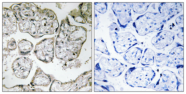
. ME2 was detected in paraffin-embedded section of human ovarian cancer tissue. Heat mediated antigen retrieval was performed in EDTA buffer (pH8.0, epitope retrieval solution). The tissue section was blocked with 10% goat serum. The tissue section was then incubated with 1microg/ml rabbit anti-ME2 Antibody (A01380-1) overnight at 4°C. Biotinylated goat anti-rabbit IgG was used as secondary antibody and incubated for 30 minutes at 37°C. The tissue section was developed using Strepavidin-Biotin-Complex (SABC) (Catalog # SA1022) with DAB as the chromogen.")
. ME2 was detected in paraffin-embedded section of human rectal cancer tissue. Heat mediated antigen retrieval was performed in EDTA buffer (pH8.0, epitope retrieval solution). The tissue section was blocked with 10% goat serum. The tissue section was then incubated with 1microg/ml rabbit anti-ME2 Antibody (A01380-1) overnight at 4°C. Biotinylated goat anti-rabbit IgG was used as secondary antibody and incubated for 30 minutes at 37°C. The tissue section was developed using Strepavidin-Biotin-Complex (SABC) (Catalog # SA1022) with DAB as the chromogen.")
. ME2 was detected in paraffin-embedded section of human rectal cancer tissue. Heat mediated antigen retrieval was performed in EDTA buffer (pH8.0, epitope retrieval solution). The tissue section was blocked with 10% goat serum. The tissue section was then incubated with 1microg/ml rabbit anti-ME2 Antibody (A01380-1) overnight at 4°C. Biotinylated goat anti-rabbit IgG was used as secondary antibody and incubated for 30 minutes at 37°C. The tissue section was developed using Strepavidin-Biotin-Complex (SABC) (Catalog # SA1022) with DAB as the chromogen.")
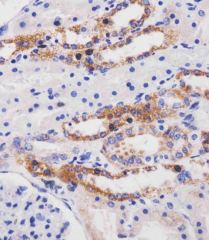
. ME2 was detected in paraffin-embedded section of rat intestine tissue. Heat mediated antigen retrieval was performed in EDTA buffer (pH8.0, epitope retrieval solution). The tissue section was blocked with 10% goat serum. The tissue section was then incubated with 1microg/ml rabbit anti-ME2 Antibody (A01380-1) overnight at 4°C. Biotinylated goat anti-rabbit IgG was used as secondary antibody and incubated for 30 minutes at 37°C. The tissue section was developed using Strepavidin-Biotin-Complex (SABC) (Catalog # SA1022) with DAB as the chromogen.")
. ME2 was detected in immunocytochemical section of Hela cells. Enzyme antigen retrieval was performed using IHC enzyme antigen retrieval reagent (AR0022) for 15 mins. The cells were blocked with 10% goat serum. And then incubated with 2microg/mL rabbit anti-ME2 Antibody (A01380-1) overnight at 4°C. DyLight®488 Conjugated Goat Anti-Rabbit IgG (BA1127) was used as secondary antibody at 1:100 dilution and incubated for 30 minutes at 37°C. The section was counterstained with DAPI. Visualize using a fluorescence microscope and filter sets appropriate for the label used.")
. ME2 was detected in paraffin-embedded section of human intestinal cancer tissue. Heat mediated antigen retrieval was performed in EDTA buffer (pH8.0, epitope retrieval solution). The tissue section was blocked with 10% goat serum. The tissue section was then incubated with 2microg/mL rabbit anti-ME2 Antibody (A01380-1) overnight at 4°C. DyLight®488 Conjugated Goat Anti-Rabbit IgG (BA1127) was used as secondary antibody at 1:100 dilution and incubated for 30 minutes at 37°C. The section was counterstained with DAPI. Visualize using a fluorescence microscope and filter sets appropriate for the label used.")
Figure 1. Western blot analysis of ME2 using anti-ME2 antibody (A01380-1). Electrophoresis was performed on a 5-20% SDS-PAGE gel at 70V (Stacking gel) / 90V (Resolving gel) for 2-3 hours. The sample well of each lane was loaded with 50ug of sample under reducing conditions. Lane 1: human Jurkat whole cell lysates, Lane 2: human K562 whole cell lysates, Lane 3: human HEK293 whole cell lysates, Lane 4: human placenta tissue lysates, Lane 5: human PC-3 whole cell lysates, Lane 6: monkey COS-7 whole cell lysates. After Electrophoresis, proteins were transferred to a Nitrocellulose membrane at 150mA for 50-90 minutes. Blocked the membrane with 5% Non-fat Milk/ TBS for 1.5 hour at RT. The membrane was incubated with rabbit anti-ME2 antigen affinity purified polyclonal antibody (Catalog # A01380-1) at 0.25 microg/mL overnight at 4°C, then washed with TBS-0.1%Tween 3 times with 5 minutes each and probed with a goat anti-rabbit IgG-HRP secondary antibody at a dilution of 1:5000 for 1.5 hour at RT. The signal is developed using an Enhanced Chemiluminescent detection (ECL) kit (Catalog # EK1002) with Tanon 5200 system. A specific band was detected for ME2 at approximately 65KD. The expected band size for ME2 is at 65KD.
Anti-ME2 Antibody Picoband(r)
A01380-1-CARRIER-FREE
ApplicationsFlow Cytometry, ImmunoFluorescence, Western Blot, ELISA, ImmunoCytoChemistry, ImmunoHistoChemistry
Product group Antibodies
ReactivityHuman, Monkey, Mouse, Rat
TargetME2
Overview
- SupplierBoster Bio
- Product NameAnti-ME2 Antibody Picoband(r)
- Delivery Days Customer9
- ApplicationsFlow Cytometry, ImmunoFluorescence, Western Blot, ELISA, ImmunoCytoChemistry, ImmunoHistoChemistry
- CertificationResearch Use Only
- ClonalityPolyclonal
- Concentration500 ug/ml
- Gene ID4200
- Target nameME2
- Target descriptionmalic enzyme 2
- Target synonymsODS1, NAD-dependent malic enzyme, mitochondrial, NAD-ME, malate dehydrogenase (oxaloacetate-decarboxylating), malic enzyme 2, NAD(+)-dependent, mitochondrial, pyruvic-malic carboxylase
- HostRabbit
- IsotypeIgG
- Protein IDP23368
- Protein NameNAD-dependent malic enzyme, mitochondrial
- Scientific DescriptionBoster Bio Anti-ME2 Antibody Picoband® catalog # A01380-1. Tested in ELISA, Flow Cytometry, IF, IHC, ICC, WB applications. This antibody reacts with Human, Monkey, Mouse, Rat. The brand Picoband indicates this is a premium antibody that guarantees superior quality, high affinity, and strong signals with minimal background in Western blot applications. Only our best-performing antibodies are designated as Picoband, ensuring unmatched performance.
- ReactivityHuman, Monkey, Mouse, Rat
- Storage Instruction-20°C,2°C to 8°C
- UNSPSC12352203
Related products
Product group Antibodies
Anti-ME2 AntibodyA100051
ApplicationsWestern Blot, ELISA, ImmunoHistoChemistry
ReactivityHuman
- SizePrice

Product group Antibodies
Anti-ME2 AntibodyHPA008247
ApplicationsWestern Blot, ImmunoCytoChemistry, ImmunoHistoChemistry
ReactivityHuman
TargetME2
- SizePrice
Product group Antibodies
ME2 AntibodyCSB-PA010151
ApplicationsWestern Blot, ELISA, ImmunoHistoChemistry
ReactivityHuman
TargetME2
- SizePrice
Product group Antibodies

ME2 / Malate Dehydrogenase 2 AntibodyLS-C411185
ApplicationsWestern Blot
ReactivityHuman, Mouse
TargetME2
- SizePrice
Product group Antibodies
ME2 Polyclonal AntibodyCAC13111
ApplicationsELISA, ImmunoHistoChemistry
TargetME2
- SizePrice
Product group Antibodies
ME2 antibodyGTX130673
ApplicationsImmunoPrecipitation, Western Blot, ImmunoHistoChemistry, ImmunoHistoChemistry Paraffin
ReactivityHuman, Mouse
TargetME2
- SizePrice
Product group Antibodies
Anti-ME2 Antibody144-09650
ApplicationsWestern Blot
ReactivityHuman, Mouse
TargetME2
- SizePrice
Product group Antibodies
ME2 Monoclonal AntibodyBSM-51670M
ApplicationsImmunoFluorescence, Western Blot, ImmunoHistoChemistry, ImmunoHistoChemistry Frozen, ImmunoHistoChemistry Paraffin
ReactivityHuman, Mouse
TargetME2
- SizePrice