


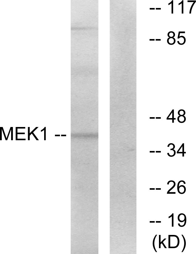
Figure 1. Western blot analysis of MEK1-2 using anti-MEK1-2 antibody (M00292-1). Electrophoresis was performed on a 5-20% SDS-PAGE gel at 70V (Stacking gel) / 90V (Resolving gel) for 2-3 hours. The sample well of each lane was loaded with 30 ug of sample under reducing conditions. Lane 1: human 293T whole cell lysates, Lane 2: human A549 whole cell lysates, Lane 3: human A431 whole cell lysates, Lane 4: human U2OS whole cell lysates, Lane 5: rat brain tissue lysates, Lane 6: rat lung tissue lysates, Lane 7: mouse brain tissue lysates, Lane 8: mouse lung tissue lysates. After electrophoresis, proteins were transferred to a nitrocellulose membrane at 150 mA for 50-90 minutes. Blocked the membrane with 5% non-fat milk/TBS for 1.5 hour at RT. The membrane was incubated with rabbit anti-MEK1-2 antigen affinity purified monoclonal antibody (Catalog # M00292-1) at 1:5000 overnight at 4°C, then washed with TBS-0.1%Tween 3 times with 5 minutes each and probed with a goat anti-rabbit IgG-HRP secondary antibody at a dilution of 1:500 for 1.5 hour at RT. The signal is developed using an Enhanced Chemiluminescent detection (ECL) kit (Catalog # EK1002) with Tanon 5200 system. A specific band was detected for MEK1-2 at approximately 43 kDa. The expected band size for MEK1-2 is at 43 kDa.


Figure 1. Western blot analysis of MEK1-2 using anti-MEK1-2 antibody (M00292-1). Electrophoresis was performed on a 5-20% SDS-PAGE gel at 70V (Stacking gel) / 90V (Resolving gel) for 2-3 hours. The sample well of each lane was loaded with 30 ug of sample under reducing conditions. Lane 1: human 293T whole cell lysates, Lane 2: human A549 whole cell lysates, Lane 3: human A431 whole cell lysates, Lane 4: human U2OS whole cell lysates, Lane 5: rat brain tissue lysates, Lane 6: rat lung tissue lysates, Lane 7: mouse brain tissue lysates, Lane 8: mouse lung tissue lysates. After electrophoresis, proteins were transferred to a nitrocellulose membrane at 150 mA for 50-90 minutes. Blocked the membrane with 5% non-fat milk/TBS for 1.5 hour at RT. The membrane was incubated with rabbit anti-MEK1-2 antigen affinity purified monoclonal antibody (Catalog # M00292-1) at 1:5000 overnight at 4°C, then washed with TBS-0.1%Tween 3 times with 5 minutes each and probed with a goat anti-rabbit IgG-HRP secondary antibody at a dilution of 1:500 for 1.5 hour at RT. The signal is developed using an Enhanced Chemiluminescent detection (ECL) kit (Catalog # EK1002) with Tanon 5200 system. A specific band was detected for MEK1-2 at approximately 43 kDa. The expected band size for MEK1-2 is at 43 kDa.
Anti-MEK1/2 MAP2K1 Rabbit Monoclonal Antibody
M00292-1
ApplicationsImmunoFluorescence, ImmunoPrecipitation, Western Blot, ImmunoCytoChemistry, ImmunoHistoChemistry
Product group Antibodies
ReactivityHuman, Mouse, Rat
TargetMAP2K1
Overview
- SupplierBoster Bio
- Product NameAnti-MEK1/2 MAP2K1 Rabbit Monoclonal Antibody
- Delivery Days Customer9
- ApplicationsImmunoFluorescence, ImmunoPrecipitation, Western Blot, ImmunoCytoChemistry, ImmunoHistoChemistry
- CertificationResearch Use Only
- ClonalityMonoclonal
- Clone IDBOA-13
- Gene ID5604
- Target nameMAP2K1
- Target descriptionmitogen-activated protein kinase kinase 1
- Target synonymsCFC3, MAPKK1, MEK1, MEL, MKK1, PRKMK1, dual specificity mitogen-activated protein kinase kinase 1, ERK activator kinase 1, MAPK/ERK kinase 1, MAPKK 1, MEK 1, protein kinase, mitogen-activated, kinase 1 (MAP kinase kinase 1)
- HostRabbit
- IsotypeIgG
- Protein IDP36507
- Protein NameDual specificity mitogen-activated protein kinase kinase 2
- Scientific DescriptionBoster Bio Anti-MEK1/2 MAP2K1 Rabbit Monoclonal Antibody catalog # M00292-1. Tested in WB, IHC, ICC/IF, IP applications. This antibody reacts with Human, Mouse, Rat.
- ReactivityHuman, Mouse, Rat
- Storage Instruction-20°C
- UNSPSC12352203
References
- Bai X, Liu X, Li S, et al. Nuciferine Inhibits TMEM16A in Dietary Adjuvant Therapy for Lung Cancer. J Agric Food Chem. 2022,70(12):3687-3696. doi: 10.1021/acs.jafc.1c08375Read this paper
Datasheet
MSDS
Related products
Product group Antibodies
Anti-MEK1 AntibodyA96159
ApplicationsWestern Blot, ELISA, ImmunoHistoChemistry
ReactivityHuman, Mouse, Rat
- SizePrice
Product group Antibodies
Anti-MAP2K1 Antibody144-00252
ApplicationsImmunoFluorescence, Western Blot, ImmunoHistoChemistry
ReactivityHuman, Mouse, Rat
TargetMAP2K1
- SizePrice

Product group Antibodies
Anti-MEK1/MAP2K1 Antibody Picoband(r)A00292-CARRIER-FREE
ApplicationsFlow Cytometry, ImmunoFluorescence, Western Blot, ELISA, ImmunoCytoChemistry
ReactivityHuman, Mouse, Rat
TargetMAP2K1
- SizePrice
Product group Antibodies
References
MEK1 + MEK2 Polyclonal AntibodyBS-1041R
ApplicationsImmunoFluorescence, Western Blot, ELISA, ImmunoCytoChemistry, ImmunoHistoChemistry, ImmunoHistoChemistry Frozen, ImmunoHistoChemistry Paraffin
TargetMAP2K1
- SizePrice
Product group Antibodies
MAP2K1 AntibodyCSB-PA003217
ApplicationsWestern Blot, ELISA, ImmunoHistoChemistry
ReactivityHuman, Mouse, Rat
TargetMAP2K1
- SizePrice
Product group Antibodies
MAP2K1 Polyclonal AntibodyCAC14738
ApplicationsWestern Blot, ELISA, ImmunoHistoChemistry
ReactivityMouse
TargetMAP2K1
- SizePrice
Product group Antibodies

MAP2K1 / MKK1 / MEK1 AntibodyLS-C405977
ApplicationsWestern Blot, ELISA, ImmunoHistoChemistry
ReactivityHuman, Mouse, Rat
TargetMAP2K1
- SizePrice
Product group Antibodies
MEK1 antibodyGTX102391
ApplicationsImmunoFluorescence, Western Blot, ImmunoCytoChemistry, ImmunoHistoChemistry, ImmunoHistoChemistry Paraffin
ReactivityCanine, Human, Mouse, Rat
TargetMAP2K1
- SizePrice

Product group Antibodies
Anti-MAP2K1 AntibodyHPA026430
ApplicationsWestern Blot, ImmunoCytoChemistry, ImmunoHistoChemistry
ReactivityHuman
TargetMAP2K1
- SizePrice