


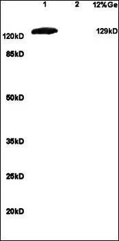
Figure 1. Western blot analysis of mGluR1/GRM1 using anti-mGluR1/GRM1 antibody (A03049-4). Electrophoresis was performed on a 5-20% SDS-PAGE gel at 70V (Stacking gel) / 90V (Resolving gel) for 2-3 hours. The sample well of each lane was loaded with 50ug of sample under reducing conditions. Lane 1: human placenta tissue lysates, Lane 2: human Hela whole cell lysates, Lane 3: human A431 whole cell lysates, Lane 4: human A549 whole cell lysates, Lane 5: human K562 whole cell lysates, Lane 6: rat brain tissue lysates, Lane 7: mouse brain tissue lysates. After Electrophoresis, proteins were transferred to a Nitrocellulose membrane at 150mA for 50-90 minutes. Blocked the membrane with 5% Non-fat Milk/ TBS for 1.5 hour at RT. The membrane was incubated with rabbit anti-mGluR1/GRM1 antigen affinity purified polyclonal antibody (Catalog # A03049-4) at 0.25 microg/mL overnight at 4°C, then washed with TBS-0.1%Tween 3 times with 5 minutes each and probed with a goat anti-rabbit IgG-HRP secondary antibody at a dilution of 1:10000 for 1.5 hour at RT. The signal is developed using an Enhanced Chemiluminescent detection (ECL) kit (Catalog # EK1002) with Tanon 5200 system. A specific band was detected for mGluR1/GRM1 at approximately 132KD. The expected band size for mGluR1/GRM1 is at 132KD.
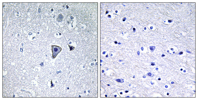
. Integrin beta 4/ITGB4 was detected in paraffin-embedded section of human glioma tissue. Heat mediated antigen retrieval was performed in EDTA buffer (pH8.0, epitope retrieval solution). The tissue section was blocked with 10% goat serum. The tissue section was then incubated with 1microg/ml rabbit anti-Integrin beta 4/ITGB4 Antibody (A03049-4) overnight at 4°C. Biotinylated goat anti-rabbit IgG was used as secondary antibody and incubated for 30 minutes at 37°C. The tissue section was developed using Strepavidin-Biotin-Complex (SABC) (Catalog # SA1022) with DAB as the chromogen.")
. Overlay histogram showing A431 cells stained with A03049-4 (Blue line). To facilitate intracellular staining, cells were fixed with 4% paraformaldehyde and permeabilized with permeabilization buffer. The cells were blocked with 10% normal goat serum. And then incubated with rabbit anti-mGluR1/GRM1 Antibody (A03049-4,1microg/1x106 cells) for 30 min at 20°C. DyLight®488 conjugated goat anti-rabbit IgG (BA1127, 5-10microg/1x106 cells) was used as secondary antibody for 30 minutes at 20°C. Isotype control antibody (Green line) was rabbit IgG (1microg/1x106) used under the same conditions. Unlabelled sample without incubation with primary antibody and secondary antibody (Red line) was used as a blank control.")
. Overlay histogram showing U20S cells stained with A03049-4 (Blue line). To facilitate intracellular staining, cells were fixed with 4% paraformaldehyde and permeabilized with permeabilization buffer. The cells were blocked with 10% normal goat serum. And then incubated with rabbit anti-mGluR1/GRM1 Antibody (A03049-4,1microg/1x106 cells) for 30 min at 20°C. DyLight®488 conjugated goat anti-rabbit IgG (BA1127, 5-10microg/1x106 cells) was used as secondary antibody for 30 minutes at 20°C. Isotype control antibody (Green line) was rabbit IgG (1microg/1x106) used under the same conditions. Unlabelled sample without incubation with primary antibody and secondary antibody (Red line) was used as a blank control.")
. Overlay histogram showing Neuro-2a cells stained with A03049-4 (Blue line). To facilitate intracellular staining, cells were fixed with 4% paraformaldehyde and permeabilized with permeabilization buffer. The cells were blocked with 10% normal goat serum. And then incubated with rabbit anti-mGluR1/GRM1 Antibody (A03049-4,1microg/1x106 cells) for 30 min at 20°C. DyLight®488 conjugated goat anti-rabbit IgG (BA1127, 5-10microg/1x106 cells) was used as secondary antibody for 30 minutes at 20°C. Isotype control antibody (Green line) was rabbit IgG (1microg/1x106) used under the same conditions. Unlabelled sample without incubation with primary antibody and secondary antibody (Red line) was used as a blank control.")
. Integrin beta 4/ITGB4 was detected in paraffin-embedded section of rat brain tissue. Heat mediated antigen retrieval was performed in EDTA buffer (pH8.0, epitope retrieval solution). The tissue section was blocked with 10% goat serum. The tissue section was then incubated with 1microg/ml rabbit anti-Integrin beta 4/ITGB4 Antibody (A03049-4) overnight at 4°C. Biotinylated goat anti-rabbit IgG was used as secondary antibody and incubated for 30 minutes at 37°C. The tissue section was developed using Strepavidin-Biotin-Complex (SABC) (Catalog # SA1022) with DAB as the chromogen.")
Figure 1. Western blot analysis of mGluR1/GRM1 using anti-mGluR1/GRM1 antibody (A03049-4). Electrophoresis was performed on a 5-20% SDS-PAGE gel at 70V (Stacking gel) / 90V (Resolving gel) for 2-3 hours. The sample well of each lane was loaded with 50ug of sample under reducing conditions. Lane 1: human placenta tissue lysates, Lane 2: human Hela whole cell lysates, Lane 3: human A431 whole cell lysates, Lane 4: human A549 whole cell lysates, Lane 5: human K562 whole cell lysates, Lane 6: rat brain tissue lysates, Lane 7: mouse brain tissue lysates. After Electrophoresis, proteins were transferred to a Nitrocellulose membrane at 150mA for 50-90 minutes. Blocked the membrane with 5% Non-fat Milk/ TBS for 1.5 hour at RT. The membrane was incubated with rabbit anti-mGluR1/GRM1 antigen affinity purified polyclonal antibody (Catalog # A03049-4) at 0.25 microg/mL overnight at 4°C, then washed with TBS-0.1%Tween 3 times with 5 minutes each and probed with a goat anti-rabbit IgG-HRP secondary antibody at a dilution of 1:10000 for 1.5 hour at RT. The signal is developed using an Enhanced Chemiluminescent detection (ECL) kit (Catalog # EK1002) with Tanon 5200 system. A specific band was detected for mGluR1/GRM1 at approximately 132KD. The expected band size for mGluR1/GRM1 is at 132KD.
Anti-mGluR1/GRM1 Antibody Picoband(r)
A03049-4-CARRIER-FREE
ApplicationsFlow Cytometry, Western Blot, ELISA, ImmunoHistoChemistry
Product group Antibodies
ReactivityHuman, Mouse, Rat
TargetGRM1
Overview
- SupplierBoster Bio
- Product NameAnti-mGluR1/GRM1 Antibody Picoband(r)
- Delivery Days Customer9
- ApplicationsFlow Cytometry, Western Blot, ELISA, ImmunoHistoChemistry
- CertificationResearch Use Only
- ClonalityPolyclonal
- Concentration500 ug/ml
- Gene ID2911
- Target nameGRM1
- Target descriptionglutamate metabotropic receptor 1
- Target synonymsGPRC1A, MGLU1, MGLUR1, PPP1R85, SCA44, SCAR13, metabotropic glutamate receptor 1, glutamate receptor, metabotropic 1, protein phosphatase 1, regulatory subunit 85
- HostRabbit
- IsotypeIgG
- Protein IDQ13255
- Protein NameMetabotropic glutamate receptor 1
- Scientific DescriptionBoster Bio Anti-mGluR1/GRM1 Antibody Picoband® catalog # A03049-4. Tested in ELISA, Flow Cytometry, IHC, WB applications. This antibody reacts with Human, Mouse, Rat. The brand Picoband indicates this is a premium antibody that guarantees superior quality, high affinity, and strong signals with minimal background in Western blot applications. Only our best-performing antibodies are designated as Picoband, ensuring unmatched performance.
- ReactivityHuman, Mouse, Rat
- Storage Instruction-20°C,2°C to 8°C
- UNSPSC12352203
Related products
Product group Antibodies
Anti-GRM1 AntibodyA97783
ApplicationsELISA, ImmunoHistoChemistry
ReactivityHuman, Mouse, Rat
- SizePrice
Product group Antibodies
Anti-GRM1 Antibody144-62066
ApplicationsWestern Blot
ReactivityHuman, Mouse, Rat
TargetGRM1
- SizePrice

Product group Antibodies
GRM1 Polyclonal AntibodyBS-1803R
ApplicationsFlow Cytometry, ImmunoFluorescence, Western Blot, ELISA, ImmunoCytoChemistry, ImmunoHistoChemistry, ImmunoHistoChemistry Frozen, ImmunoHistoChemistry Paraffin
ReactivityBovine, Canine, Chicken, Equine, Human, Mouse, Porcine, Rat
TargetGRM1
- SizePrice
Product group Antibodies
GRM1 AntibodyCSB-PA009017
ApplicationsELISA, ImmunoHistoChemistry
ReactivityHuman, Mouse, Rat
TargetGRM1
- SizePrice
Product group Antibodies
ApplicationsImmunoPrecipitation, Western Blot, ImmunoCytoChemistry, ImmunoHistoChemistry
ReactivityMouse, Porcine, Rat
TargetGRM1
- SizePrice
Product group Antibodies
mGluR1 antibodyGTX37416
ApplicationsWestern Blot, ImmunoHistoChemistry, ImmunoHistoChemistry Paraffin
ReactivityHuman, Mouse, Rat
TargetGRM1
- SizePrice

Product group Antibodies
Anti-GRM1 AntibodyHPA015701
ApplicationsImmunoHistoChemistry
ReactivityHuman
TargetGRM1
- SizePrice
Product group Antibodies

GRM1 / MGLUR1 AntibodyLS-C746882
ApplicationsWestern Blot
ReactivityHuman, Rat
TargetGRM1
- SizePrice