


Figure 2. IF analysis of NR3C2 using anti-NR3C2 antibody (PB9765) and anti-Tubulin Alpha antibody (M03989-3). NR3C2 was detected in immunocytochemical section of U20S cell. Enzyme antigen retrieval was performed using IHC enzyme antigen retrieval reagent (AR0022) for 15 mins. The cells were blocked with 10% goat serum. And then incubated with 5 microg/mL rabbit anti-NR3C2 Antibody (PB9765) and mouse anti-Tubulin Alpha antibody (M03989-3) overnight at 4°C. Cy3 Conjugated Goat Anti-Rabbit IgG (BA1032) and DyLight®488 Conjugated Goat Anti-Mouse IgG (BA1126) were used as secondary antibody at 1:500 dilution and incubated for 30 minutes at 37°C. Visualize using a fluorescence microscope and filter sets appropriate for the label used.
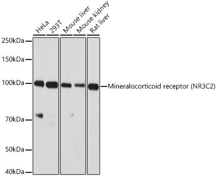
. Electrophoresis was performed on a 5-20% SDS-PAGE gel at 70V (Stacking gel) / 90V (Resolving gel) for 2-3 hours. The sample well of each lane was loaded with 30 ug of sample under reducing conditions. Lane 1: human Hela whole cell lysates, Lane 2: human 293T whole cell lysates, Lane 3: human LNCAP whole cell lysates, Lane 4: human MCF-7 whole cell lysates, Lane 5: rat NRK whole cell lysates, Lane 6: mouse NIH/3T3 whole cell lysates. After electrophoresis, proteins were transferred to a nitrocellulose membrane at 150 mA for 50-90 minutes. Blocked the membrane with 5% non-fat milk/TBS for 1.5 hour at RT. The membrane was incubated with rabbit anti-NR3C2 antigen affinity purified polyclonal antibody (Catalog # PB9765) at 0.5 microg/mL overnight at 4°C, then washed with TBS-0.1%Tween 3 times with 5 minutes each and probed with a goat anti-rabbit IgG-HRP secondary antibody at a dilution of 1:5000 for 1.5 hour at RT. The signal is developed using an Enhanced Chemiluminescent detection (ECL) kit (Catalog # EK1002) with Tanon 5200 system. A specific band was detected for NR3C2 at approximately 110 kDa. The expected band size for NR3C2 is at 107 kDa.")
Figure 2. IF analysis of NR3C2 using anti-NR3C2 antibody (PB9765) and anti-Tubulin Alpha antibody (M03989-3). NR3C2 was detected in immunocytochemical section of U20S cell. Enzyme antigen retrieval was performed using IHC enzyme antigen retrieval reagent (AR0022) for 15 mins. The cells were blocked with 10% goat serum. And then incubated with 5 microg/mL rabbit anti-NR3C2 Antibody (PB9765) and mouse anti-Tubulin Alpha antibody (M03989-3) overnight at 4°C. Cy3 Conjugated Goat Anti-Rabbit IgG (BA1032) and DyLight®488 Conjugated Goat Anti-Mouse IgG (BA1126) were used as secondary antibody at 1:500 dilution and incubated for 30 minutes at 37°C. Visualize using a fluorescence microscope and filter sets appropriate for the label used.
Anti-Mineralocorticoid Receptor/NR3C2 Antibody Picoband(r)
PB9765-CARRIER-FREE
ApplicationsImmunoFluorescence, Western Blot, ImmunoCytoChemistry
Product group Antibodies
ReactivityChicken, Human, Mouse, Rat
TargetNR3C2
Overview
- SupplierBoster Bio
- Product NameAnti-Mineralocorticoid Receptor/NR3C2 Antibody Picoband(r)
- Delivery Days Customer9
- Application Supplier NoteTested Species: In-house tested species with positive results. Other applications have not been tested. Optimal dilutions should be determined by end users.
- ApplicationsImmunoFluorescence, Western Blot, ImmunoCytoChemistry
- CertificationResearch Use Only
- ClonalityPolyclonal
- Concentration500 ug/ml
- Gene ID4306
- Target nameNR3C2
- Target descriptionnuclear receptor subfamily 3 group C member 2
- Target synonymsMCR, MLR, MR, NR3C2VIT, mineralocorticoid receptor, aldosterone receptor, mineralocorticoid receptor 1, mineralocorticoid receptor 2, mineralocorticoid receptor delta
- HostRabbit
- IsotypeIgG
- Protein IDP08235
- Protein NameMineralocorticoid receptor
- Scientific DescriptionBoster Bio Anti-Mineralocorticoid Receptor/NR3C2 Antibody Picoband® catalog # PB9765. Tested in IF, ICC, WB applications. This antibody reacts with Human, Mouse, Rat. The brand Picoband indicates this is a premium antibody that guarantees superior quality, high affinity, and strong signals with minimal background in Western blot applications. Only our best-performing antibodies are designated as Picoband, ensuring unmatched performance.
- ReactivityChicken, Human, Mouse, Rat
- Storage Instruction-20°C,2°C to 8°C
- UNSPSC12352203
Related products
Product group Antibodies
ApplicationsImmunoFluorescence, Western Blot, ImmunoCytoChemistry, ImmunoHistoChemistry
ReactivityHuman, Mouse, Rat
- SizePrice
Product group Antibodies
Anti-NR3C2 Antibody144-65691
ApplicationsWestern Blot
ReactivityHuman
TargetNR3C2
- SizePrice
Product group Antibodies
References
ApplicationsFlow Cytometry, ImmunoFluorescence, Western Blot, ELISA, ImmunoCytoChemistry, ImmunoHistoChemistry, ImmunoHistoChemistry Frozen, ImmunoHistoChemistry Paraffin
ReactivityBovine, Canine, Chicken, Human, Mouse, Porcine, Rabbit, Rat
TargetNR3C2
- SizePrice
Product group Antibodies
ApplicationsWestern Blot, ELISA, ImmunoCytoChemistry, ImmunoHistoChemistry, ImmunoHistoChemistry Frozen, ImmunoHistoChemistry Paraffin
ReactivityMouse, Porcine
TargetNR3C2
- SizePrice
Product group Antibodies
NR3C2 AntibodyCSB-PA016061GA01HU
ApplicationsImmunoFluorescence, Western Blot, ELISA
ReactivityHuman, Mouse, Rat
TargetNR3C2
- SizePrice
![ICC/IF analysis of HEK293 cells using GTX22774 Mineralocorticoid Receptor antibody [H10E4C9F]. Cells were probed without (right) or with(left) an antibody. Green : Primary antibody Blue : Nuclei Red : Actin Fixation : formaldehyde Dilution : 1:200 overnight at 4oC](https://www.genetex.com/upload/website/prouct_img/normal/GTX22774/GTX22774_426_ICC-IF_w_23060620_642.webp)
Product group Antibodies
ApplicationsFlow Cytometry, ImmunoFluorescence, ImmunoPrecipitation, Western Blot, ImmunoCytoChemistry, ImmunoHistoChemistry, ImmunoHistoChemistry Paraffin, Neutralisation/Blocking
ReactivityChicken, Human, Mouse, Rabbit, Rat, Sheep
TargetNR3C2
- SizePrice
Product group Antibodies

Mineralocorticoid Receptor AntibodyLS-C332476
ApplicationsWestern Blot
ReactivityHuman
TargetNR3C2
- SizePrice

Product group Antibodies
Anti-NR3C2 AntibodyHPA074979
ApplicationsImmunoCytoChemistry
ReactivityHuman
TargetNR3C2
- SizePrice
Product group Antibodies
Anti-NR3C2 AntibodyCAB3308
ApplicationsImmunoFluorescence, Western Blot, ELISA, ImmunoCytoChemistry, ImmunoHistoChemistry, ImmunoHistoChemistry Paraffin
ReactivityHuman
TargetNR3C2
- SizePrice