


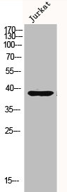
Figure 1. Western blot analysis of MKP-1/DUSP1 using anti-MKP-1/DUSP1 antibody (A02276-1). Electrophoresis was performed on a 5-20% SDS-PAGE gel at 70V (Stacking gel) / 90V (Resolving gel) for 2-3 hours. The sample well of each lane was loaded with 50ug of sample under reducing conditions. Lane 1: human U87 whole cell lysates, Lane 2: human HELA whole cell lysates, Lane 3: human A549 whole cell lysates. After Electrophoresis, proteins were transferred to a Nitrocellulose membrane at 150mA for 50-90 minutes. Blocked the membrane with 5% Non-fat Milk/ TBS for 1.5 hour at RT. The membrane was incubated with rabbit anti-MKP-1/DUSP1 antigen affinity purified polyclonal antibody (Catalog # A02276-1) at 0.5 microg/mL overnight at 4°C, then washed with TBS-0.1%Tween 3 times with 5 minutes each and probed with a goat anti-rabbit IgG-HRP secondary antibody at a dilution of 1:5000 for 1.5 hour at RT. The signal is developed using an Enhanced Chemiluminescent detection (ECL) kit (Catalog # EK1002) with Tanon 5200 system. A specific band was detected for MKP-1/DUSP1 at approximately 40KD. The expected band size for MKP-1/DUSP1 is at 40KD.
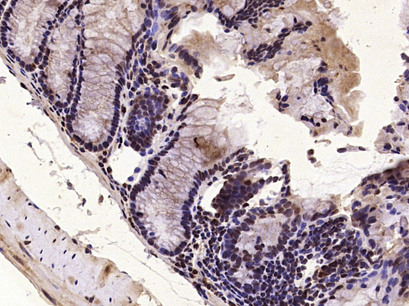
. Overlay histogram showing CACO-2 cells stained with A02276-1 (Blue line). To facilitate intracellular staining, cells were fixed with 4% paraformaldehyde and permeabilized with permeabilization buffer. The cells were blocked with 10% normal goat serum. And then incubated with rabbit anti-MKP-1/DUSP1 Antibody (A02276-1, 1microg/1x106 cells) for 30 min at 20°C. DyLight®488 conjugated goat anti-rabbit IgG (BA1127, 5-10microg/1x106 cells) was used as secondary antibody for 30 minutes at 20°C. Isotype control antibody (Green line) was rabbit IgG (1microg/1x106) used under the same conditions. Unlabelled sample without incubation with primary antibody and secondary antibody (Red line) was used as a blank control.")
Figure 1. Western blot analysis of MKP-1/DUSP1 using anti-MKP-1/DUSP1 antibody (A02276-1). Electrophoresis was performed on a 5-20% SDS-PAGE gel at 70V (Stacking gel) / 90V (Resolving gel) for 2-3 hours. The sample well of each lane was loaded with 50ug of sample under reducing conditions. Lane 1: human U87 whole cell lysates, Lane 2: human HELA whole cell lysates, Lane 3: human A549 whole cell lysates. After Electrophoresis, proteins were transferred to a Nitrocellulose membrane at 150mA for 50-90 minutes. Blocked the membrane with 5% Non-fat Milk/ TBS for 1.5 hour at RT. The membrane was incubated with rabbit anti-MKP-1/DUSP1 antigen affinity purified polyclonal antibody (Catalog # A02276-1) at 0.5 microg/mL overnight at 4°C, then washed with TBS-0.1%Tween 3 times with 5 minutes each and probed with a goat anti-rabbit IgG-HRP secondary antibody at a dilution of 1:5000 for 1.5 hour at RT. The signal is developed using an Enhanced Chemiluminescent detection (ECL) kit (Catalog # EK1002) with Tanon 5200 system. A specific band was detected for MKP-1/DUSP1 at approximately 40KD. The expected band size for MKP-1/DUSP1 is at 40KD.
Anti-MKP-1/DUSP1 Antibody Picoband(r)
A02276-1-CARRIER-FREE
ApplicationsFlow Cytometry, Western Blot
Product group Antibodies
ReactivityHuman
TargetDUSP1
Overview
- SupplierBoster Bio
- Product NameAnti-MKP-1/DUSP1 Antibody Picoband(r)
- Delivery Days Customer9
- ApplicationsFlow Cytometry, Western Blot
- CertificationResearch Use Only
- ClonalityPolyclonal
- Concentration500 ug/ml
- Gene ID1843
- Target nameDUSP1
- Target descriptiondual specificity phosphatase 1
- Target synonymsCL100, HVH1, MKP-1, MKP1, PTPN10, dual specificity protein phosphatase 1, CL 100, MAP kinase phosphatase 1, dual specificity protein phosphatase hVH1, mitogen-activated protein kinase phosphatase 1, protein-tyrosine phosphatase CL100, serine/threonine specific protein phosphatase
- HostRabbit
- IsotypeIgG
- Protein IDP28562
- Protein NameDual specificity protein phosphatase 1
- Scientific DescriptionBoster Bio Anti-MKP-1/DUSP1 Antibody Picoband® catalog # A02276-1. Tested in Flow Cytometry, WB applications. This antibody reacts with Human. The brand Picoband indicates this is a premium antibody that guarantees superior quality, high affinity, and strong signals with minimal background in Western blot applications. Only our best-performing antibodies are designated as Picoband, ensuring unmatched performance.
- ReactivityHuman
- Storage Instruction-20°C,2°C to 8°C
- UNSPSC12352203
Related products
Product group Antibodies
Anti-DUSP1 Antibody144-02919
ApplicationsWestern Blot
ReactivityHuman, Mouse
TargetDUSP1
- SizePrice

Product group Antibodies
Anti-DUSP1 AntibodyA43339
ApplicationsWestern Blot
ReactivityHuman, Mouse, Rat
- SizePrice
Product group Antibodies
References
MKP1 Polyclonal AntibodyBS-1851R
ApplicationsImmunoFluorescence, Western Blot, ELISA, ImmunoCytoChemistry, ImmunoHistoChemistry, ImmunoHistoChemistry Frozen, ImmunoHistoChemistry Paraffin
ReactivityBovine, Canine, Equine, Human, Mouse, Rabbit, Rat, Sheep
TargetDUSP1
- SizePrice
Product group Antibodies
DUSP1 AntibodyCSB-PA003245
ApplicationsWestern Blot, ELISA, ImmunoHistoChemistry
ReactivityHuman
TargetDUSP1
- SizePrice
Product group Antibodies

Goat anti-DUSP1EB05197
ApplicationsWestern Blot, ELISA, ImmunoHistoChemistry
ReactivityBovine, Canine, Human, Mouse, Rat
TargetDUSP1
- SizePrice
Product group Antibodies
DUSP1 Polyclonal AntibodyCAC15043
ApplicationsWestern Blot, ELISA
TargetDUSP1
- SizePrice
![Mouse tissue extract (50 μg) was separated by 10% SDS-PAGE, and the membranes were blotted with DUSP1 antibody [N3C3] (GTX101229) diluted at 1:1000 and competitor's antibody (sc-1199) diluted at 1:200. The HRP-conjugated anti-rabbit IgG antibody (GTX213110-01) was used to detect the primary antibody.](https://www.genetex.com/upload/website/prouct_img/normal/GTX101229/GTX101229_40128_20170810_WB_M_liver_competitor_watermark_w_23060100_718.webp)
Product group Antibodies
DUSP1 antibodyGTX101229
ApplicationsWestern Blot
ReactivityHuman, Mouse
TargetDUSP1
- SizePrice

Product group Antibodies
Anti-DUSP1 AntibodyHPA069577
ApplicationsWestern Blot, ImmunoCytoChemistry
ReactivityHuman
TargetDUSP1
- SizePrice