


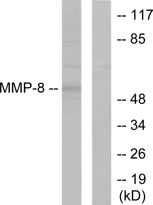
Figure 1. Western blot analysis of MMP8 using anti-MMP8 antibody (PB9726). Electrophoresis was performed on a 5-20% SDS-PAGE gel at 70V (Stacking gel) / 90V (Resolving gel) for 2-3 hours. The sample well of each lane was loaded with 40ug of sample under reducing conditions. Lane 1: K562 Whole Cell Lysate, Lane 2: JURKAT Whole Cell Lysate. After Electrophoresis, proteins were transferred to a Nitrocellulose membrane at 150mA for 50-90 minutes. Blocked the membrane with 5% Non-fat Milk/ TBS for 1.5 hour at RT. The membrane was incubated with rabbit anti-MMP8 antigen affinity purified polyclonal antibody (Catalog # PB9726) at 0.5 microg/mL overnight at 4°C, then washed with TBS-0.1%Tween 3 times with 5 minutes each and probed with a goat anti-rabbit IgG-HRP secondary antibody at a dilution of 1:10000 for 1.5 hour at RT. The signal is developed using an Enhanced Chemiluminescent detection (ECL) kit (Catalog # EK1002) with Tanon 5200 system. A specific band was detected for MMP8 at approximately 60KD. The expected band size for MMP8 is at 53KD.
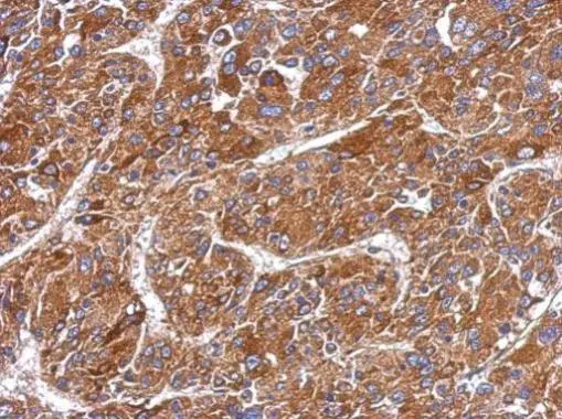
. MMP8 was detected in paraffin-embedded section of Human Appendicitis Tissue. Heat mediated antigen retrieval was performed in EDTA buffer (pH 8.0, epitope retrieval solution). The tissue section was blocked with 10% goat serum. The tissue section was then incubated with 1microg/ml rabbit anti-MMP8 Antibody (PB9726) overnight at 4°C. Biotinylated goat anti-rabbit IgG was used as secondary antibody and incubated for 30 minutes at 37°C. The tissue section was developed using Strepavidin-Biotin-Complex (SABC)(Catalog # SA1022) with DAB as the chromogen.")
Figure 1. Western blot analysis of MMP8 using anti-MMP8 antibody (PB9726). Electrophoresis was performed on a 5-20% SDS-PAGE gel at 70V (Stacking gel) / 90V (Resolving gel) for 2-3 hours. The sample well of each lane was loaded with 40ug of sample under reducing conditions. Lane 1: K562 Whole Cell Lysate, Lane 2: JURKAT Whole Cell Lysate. After Electrophoresis, proteins were transferred to a Nitrocellulose membrane at 150mA for 50-90 minutes. Blocked the membrane with 5% Non-fat Milk/ TBS for 1.5 hour at RT. The membrane was incubated with rabbit anti-MMP8 antigen affinity purified polyclonal antibody (Catalog # PB9726) at 0.5 microg/mL overnight at 4°C, then washed with TBS-0.1%Tween 3 times with 5 minutes each and probed with a goat anti-rabbit IgG-HRP secondary antibody at a dilution of 1:10000 for 1.5 hour at RT. The signal is developed using an Enhanced Chemiluminescent detection (ECL) kit (Catalog # EK1002) with Tanon 5200 system. A specific band was detected for MMP8 at approximately 60KD. The expected band size for MMP8 is at 53KD.
Anti-MMP8 Antibody Picoband(r)
PB9726-CARRIER-FREE
ApplicationsWestern Blot, ELISA, ImmunoHistoChemistry
Product group Antibodies
ReactivityHuman
TargetMMP8
Overview
- SupplierBoster Bio
- Product NameAnti-MMP8 Antibody Picoband(r)
- Delivery Days Customer9
- Application Supplier NoteTested Species: In-house tested species with positive results. By Heat: Boiling the paraffin sections in 10mM citrate buffer, pH6.0, for 20mins is required for the staining of formalin/paraffin sections. Other applications have not been tested. Optimal dilutions should be determined by end users.
- ApplicationsWestern Blot, ELISA, ImmunoHistoChemistry
- CertificationResearch Use Only
- ClonalityPolyclonal
- Concentration500 ug/ml
- Gene ID4317
- Target nameMMP8
- Target descriptionmatrix metallopeptidase 8
- Target synonymsCLG1, HNC, MMP-8, PMNL-CL, neutrophil collagenase, PMN leukocyte collagenase, PMNL collagenase, collagenase 2, matrix metalloproteinase 8 (neutrophil collagenase), matrix metalloproteinase-8
- HostRabbit
- IsotypeIgG
- Protein IDP22894
- Protein NameNeutrophil collagenase
- Scientific DescriptionBoster Bio Anti-MMP8 Antibody Picoband® catalog # PB9726. Tested in ELISA, IHC, WB applications. This antibody reacts with Human. The brand Picoband indicates this is a premium antibody that guarantees superior quality, high affinity, and strong signals with minimal background in Western blot applications. Only our best-performing antibodies are designated as Picoband, ensuring unmatched performance.
- ReactivityHuman
- Storage Instruction-20°C,2°C to 8°C
- UNSPSC12352203
Related products
Product group Antibodies
Anti-MMP-8 AntibodyA95836
ApplicationsWestern Blot, ELISA, ImmunoHistoChemistry
ReactivityHuman, Mouse, Rat
- SizePrice
Product group Antibodies
Anti-MMP8 Antibody144-01963
ApplicationsImmunoFluorescence, Western Blot
ReactivityHuman, Mouse
TargetMMP8
- SizePrice
Product group Antibodies
References
MMP8 Polyclonal AntibodyBS-1913R
ApplicationsWestern Blot, ELISA
ReactivityHuman, Mouse, Rat
TargetMMP8
- SizePrice
Product group Antibodies
MMP8 AntibodyCSB-PA010208
ApplicationsWestern Blot, ELISA, ImmunoHistoChemistry
ReactivityHuman, Mouse, Rat
TargetMMP8
- SizePrice
Product group Antibodies
MMP8 Polyclonal AntibodyCAC14654
ApplicationsImmunoFluorescence, Western Blot, ELISA
ReactivityMouse
TargetMMP8
- SizePrice
Product group Antibodies

MMP8 AntibodyLS-C405721
ApplicationsWestern Blot, ELISA, ImmunoHistoChemistry
ReactivityHuman
TargetMMP8
- SizePrice

Product group Antibodies
Anti-MMP8 AntibodyHPA021221
ApplicationsImmunoHistoChemistry
ReactivityHuman
TargetMMP8
- SizePrice
Product group Antibodies
MMP8 antibodyGTX105428
ApplicationsImmunoFluorescence, ImmunoPrecipitation, Western Blot, ImmunoCytoChemistry, ImmunoHistoChemistry, ImmunoHistoChemistry Paraffin
ReactivityHuman
TargetMMP8
- SizePrice
Product group Antibodies
ApplicationsWestern Blot, ELISA, ImmunoHistoChemistry, ImmunoHistoChemistry Paraffin
ReactivityHuman
TargetMMP8
- SizePrice