


Figure 1. Western blot analysis of MMS19 using anti-MMS19 antibody (A06218-2). Electrophoresis was performed on a 5-20% SDS-PAGE gel at 70V (Stacking gel) / 90V (Resolving gel) for 2-3 hours. The sample well of each lane was loaded with 30 ug of sample under reducing conditions. Lane 1: human Caco-2 whole cell lysates, Lane 2: mouse brain tissue lysates. After electrophoresis, proteins were transferred to a nitrocellulose membrane at 150 mA for 50-90 minutes. Blocked the membrane with 5% non-fat milk/TBS for 1.5 hour at RT. The membrane was incubated with rabbit anti-MMS19 antigen affinity purified polyclonal antibody (Catalog # A06218-2) at 0.5 microg/mL overnight at 4°C, then washed with TBS-0.1%Tween 3 times with 5 minutes each and probed with a goat anti-rabbit IgG-HRP secondary antibody at a dilution of 1:5000 for 1.5 hour at RT. The signal is developed using an Enhanced Chemiluminescent detection (ECL) kit (Catalog # EK1002) with Tanon 5200 system. A specific band was detected for MMS19 at approximately 113 kDa. The expected band size for MMS19 is at 113 kDa.
. MMS19 was detected in a paraffin-embedded section of human bladder urothelial carcinoma tissue. Heat mediated antigen retrieval was performed in EDTA buffer (pH 8.0, epitope retrieval solution). The tissue section was blocked with 10% goat serum. The tissue section was then incubated with 2 microg/ml rabbit anti-MMS19 Antibody (A06218-2) overnight at 4°C. Peroxidase Conjugated Goat Anti-rabbit IgG was used as secondary antibody and incubated for 30 minutes at 37°C. The tissue section was developed using HRP Conjugated Rabbit IgG Super Vision Assay Kit (Catalog # SV0002) with DAB as the chromogen.")
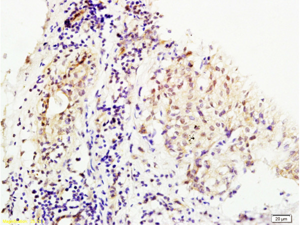
. MMS19 was detected in a paraffin-embedded section of human colorectal adenocarcinoma tissue. Heat mediated antigen retrieval was performed in EDTA buffer (pH 8.0, epitope retrieval solution). The tissue section was blocked with 10% goat serum. The tissue section was then incubated with 2 microg/ml rabbit anti-MMS19 Antibody (A06218-2) overnight at 4°C. Peroxidase Conjugated Goat Anti-rabbit IgG was used as secondary antibody and incubated for 30 minutes at 37°C. The tissue section was developed using HRP Conjugated Rabbit IgG Super Vision Assay Kit (Catalog # SV0002) with DAB as the chromogen.")
. MMS19 was detected in a paraffin-embedded section of human placenta tissue. Heat mediated antigen retrieval was performed in EDTA buffer (pH 8.0, epitope retrieval solution). The tissue section was blocked with 10% goat serum. The tissue section was then incubated with 2 microg/ml rabbit anti-MMS19 Antibody (A06218-2) overnight at 4°C. Peroxidase Conjugated Goat Anti-rabbit IgG was used as secondary antibody and incubated for 30 minutes at 37°C. The tissue section was developed using HRP Conjugated Rabbit IgG Super Vision Assay Kit (Catalog # SV0002) with DAB as the chromogen.")
. MMS19 was detected in an immunocytochemical section of HELA cells. Enzyme antigen retrieval was performed using IHC enzyme antigen retrieval reagent (AR0022) for 15 mins. The cells were blocked with 10% goat serum. And then incubated with 5 microg/mL rabbit anti-MMS19 Antibody (A06218-2) overnight at 4°C. Cy3 Conjugated Goat Anti-Rabbit IgG (BA1032) was used as secondary antibody at 1:500 dilution and incubated for 30 minutes at 37°C. Visualize using a fluorescence microscope and filter sets appropriate for the label used.")
. Overlay histogram showing Hela cells stained with A06218-2 (Blue line). To facilitate intracellular staining, cells were fixed with 4% paraformaldehyde and permeabilized with permeabilization buffer. The cells were blocked with 10% normal goat serum. And then incubated with rabbit anti-MMS19 Antibody (A06218-2, 1 microg/1x106 cells) for 30 min at 20°C. DyLight®488 conjugated goat anti-rabbit IgG (BA1127, 5-10 microg/1x106 cells) was used as secondary antibody for 30 minutes at 20°C. Isotype control antibody (Green line) was rabbit IgG (1 microg/1x106) used under the same conditions. Unlabelled sample (Red line) was also used as a control.")
Figure 1. Western blot analysis of MMS19 using anti-MMS19 antibody (A06218-2). Electrophoresis was performed on a 5-20% SDS-PAGE gel at 70V (Stacking gel) / 90V (Resolving gel) for 2-3 hours. The sample well of each lane was loaded with 30 ug of sample under reducing conditions. Lane 1: human Caco-2 whole cell lysates, Lane 2: mouse brain tissue lysates. After electrophoresis, proteins were transferred to a nitrocellulose membrane at 150 mA for 50-90 minutes. Blocked the membrane with 5% non-fat milk/TBS for 1.5 hour at RT. The membrane was incubated with rabbit anti-MMS19 antigen affinity purified polyclonal antibody (Catalog # A06218-2) at 0.5 microg/mL overnight at 4°C, then washed with TBS-0.1%Tween 3 times with 5 minutes each and probed with a goat anti-rabbit IgG-HRP secondary antibody at a dilution of 1:5000 for 1.5 hour at RT. The signal is developed using an Enhanced Chemiluminescent detection (ECL) kit (Catalog # EK1002) with Tanon 5200 system. A specific band was detected for MMS19 at approximately 113 kDa. The expected band size for MMS19 is at 113 kDa.
Anti-MMS19 Antibody Picoband(r)
A06218-2-CARRIER-FREE
ApplicationsFlow Cytometry, ImmunoFluorescence, Western Blot, ELISA, ImmunoCytoChemistry, ImmunoHistoChemistry
Product group Antibodies
ReactivityHuman, Mouse
TargetMMS19
Overview
- SupplierBoster Bio
- Product NameAnti-MMS19 Antibody Picoband(r)
- Delivery Days Customer9
- ApplicationsFlow Cytometry, ImmunoFluorescence, Western Blot, ELISA, ImmunoCytoChemistry, ImmunoHistoChemistry
- CertificationResearch Use Only
- ClonalityPolyclonal
- Concentration500 ug/ml
- Gene ID64210
- Target nameMMS19
- Target descriptionMMS19 cytosolic iron-sulfur assembly component
- Target synonymsCIAO4, MET18, MMS19L, hMMS19, MMS19 nucleotide excision repair protein homolog, MET18 homolog, MMS19 homolog, cytosolic iron-sulfur assembly component, MMS19 nucleotide excision repair homolog, MMS19-like (MET18 homolog, S. cerevisiae), MMS19-like protein, homolog of yeast MMS19
- HostRabbit
- IsotypeIgG
- Protein IDQ96T76
- Protein NameMMS19 nucleotide excision repair protein homolog
- Scientific DescriptionBoster Bio Anti-MMS19 Antibody Picoband® catalog # A06218-2. Tested in ELISA, IF, IHC, ICC, WB, Flow Cytometry applications. This antibody reacts with Human, Mouse. The brand Picoband indicates this is a premium antibody that guarantees superior quality, high affinity, and strong signals with minimal background in Western blot applications. Only our best-performing antibodies are designated as Picoband, ensuring unmatched performance.
- ReactivityHuman, Mouse
- Storage Instruction-20°C,2°C to 8°C
- UNSPSC12352203
Related products
Product group Antibodies
MMS19 Polyclonal AntibodyBS-8671R
ApplicationsImmunoFluorescence, Western Blot, ELISA, ImmunoCytoChemistry, ImmunoHistoChemistry, ImmunoHistoChemistry Frozen, ImmunoHistoChemistry Paraffin
ReactivityCanine, Equine, Human, Mouse, Porcine, Rabbit, Rat
TargetMMS19
- SizePrice
Product group Antibodies
Anti-MMS19 Antibody144-65848
ApplicationsWestern Blot
ReactivityHuman, Mouse
TargetMMS19
- SizePrice
Product group Antibodies

MMS19 AntibodyLS-C834249
ApplicationsWestern Blot
ReactivityHuman, Mouse
TargetMMS19
- SizePrice

Product group Antibodies
Anti-MMS19 AntibodyHPA051936
ApplicationsImmunoCytoChemistry
ReactivityHuman
TargetMMS19
- SizePrice
Product group Antibodies
MMS19 AntibodyCSB-PA014682GA01HU
ApplicationsWestern Blot, ELISA
ReactivityHuman, Mouse, Rat
TargetMMS19
- SizePrice