


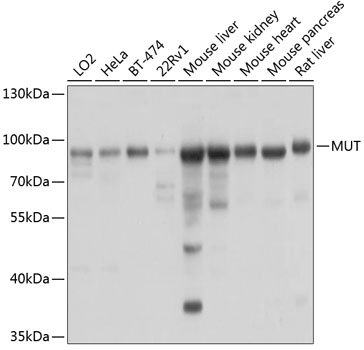
Figure 1. Western blot analysis of MMUT using anti-MMUT antibody (M34008). Electrophoresis was performed on a 5-20% SDS-PAGE gel at 70V (Stacking gel) / 90V (Resolving gel) for 2-3 hours. The sample well of each lane was loaded with 30 ug of sample under reducing conditions. Lane 1: human K562 whole cell lysates, Lane 2: human 293T whole cell lysates, Lane 3: human PC-3 whole cell lysates, Lane 4: rat PC-12 whole cell lysates, Lane 5: rat kidney tissue lysates, Lane 6: mouse NIH/3T3 whole cell lysates, Lane 7: mouse kidney tissue lysates. After electrophoresis, proteins were transferred to a nitrocellulose membrane at 150 mA for 50-90 minutes. Blocked the membrane with 5% non-fat milk/TBS for 1.5 hour at RT. The membrane was incubated with rabbit anti-MMUT antigen affinity purified monoclonal antibody (Catalog # M34008) at 1:500 overnight at 4°C, then washed with TBS-0.1%Tween 3 times with 5 minutes each and probed with a goat anti-rabbit IgG-HRP secondary antibody at a dilution of 1:500 for 1.5 hour at RT. The signal is developed using an Enhanced Chemiluminescent detection (ECL) kit (Catalog # EK1002) with Tanon 5200 system. A specific band was detected for MMUT at approximately 83 kDa. The expected band size for MMUT is at 83 kDa.
Figure 1. Western blot analysis of MMUT using anti-MMUT antibody (M34008). Electrophoresis was performed on a 5-20% SDS-PAGE gel at 70V (Stacking gel) / 90V (Resolving gel) for 2-3 hours. The sample well of each lane was loaded with 30 ug of sample under reducing conditions. Lane 1: human K562 whole cell lysates, Lane 2: human 293T whole cell lysates, Lane 3: human PC-3 whole cell lysates, Lane 4: rat PC-12 whole cell lysates, Lane 5: rat kidney tissue lysates, Lane 6: mouse NIH/3T3 whole cell lysates, Lane 7: mouse kidney tissue lysates. After electrophoresis, proteins were transferred to a nitrocellulose membrane at 150 mA for 50-90 minutes. Blocked the membrane with 5% non-fat milk/TBS for 1.5 hour at RT. The membrane was incubated with rabbit anti-MMUT antigen affinity purified monoclonal antibody (Catalog # M34008) at 1:500 overnight at 4°C, then washed with TBS-0.1%Tween 3 times with 5 minutes each and probed with a goat anti-rabbit IgG-HRP secondary antibody at a dilution of 1:500 for 1.5 hour at RT. The signal is developed using an Enhanced Chemiluminescent detection (ECL) kit (Catalog # EK1002) with Tanon 5200 system. A specific band was detected for MMUT at approximately 83 kDa. The expected band size for MMUT is at 83 kDa.
Anti-MUTA Rabbit Monoclonal Antibody
M34008
ApplicationsImmunoFluorescence, Western Blot, ImmunoCytoChemistry, ImmunoHistoChemistry
Product group Antibodies
ReactivityHuman, Mouse, Rat
TargetMMUT
Overview
- SupplierBoster Bio
- Product NameAnti-MUTA Rabbit Monoclonal Antibody
- Delivery Days Customer9
- ApplicationsImmunoFluorescence, Western Blot, ImmunoCytoChemistry, ImmunoHistoChemistry
- CertificationResearch Use Only
- ClonalityMonoclonal
- Clone ID27M55
- Gene ID4594
- Target nameMMUT
- Target descriptionmethylmalonyl-CoA mutase
- Target synonymsMCM, MUT, methylmalonyl-CoA mutase, mitochondrial, methylmalonyl Coenzyme A mutase, methylmalonyl-CoA isomerase, methylmalonyl-CoA mutase c.*192delA, methylmalonyl-CoA mutase c.*51C>G, methylmalonyl-CoA mutase variant c.1495G>A, methylmalonyl-CoA mutase variant c.2011A>G, methylmalonyl-CoA mutase variant c.2150G>T, methylmalonyl-CoA mutase variant c.322C>T, methylmalonyl-CoA mutase variant c.613_615delGAA, methylmalonyl-CoA mutase variant c.636G>A, methylmalonyl-CoA mutase variant c.643G>A, mutant methylmalonyl CoA mutase
- HostRabbit
- IsotypeIgG
- Protein IDP22033
- Protein NameMethylmalonyl-CoA mutase, mitochondrial
- Scientific DescriptionBoster Bio Anti-MUTA Rabbit Monoclonal Antibody catalog # M34008. Tested in WB, IHC, ICC/IF applications. This antibody reacts with Human, Mouse, Rat.
- ReactivityHuman, Mouse, Rat
- Storage Instruction-20°C
- UNSPSC12352203
Related products
Product group Antibodies
ApplicationsWestern Blot, ImmunoCytoChemistry
ReactivityHuman, Mouse, Rat
TargetMMUT
- SizePrice
Product group Antibodies
MMUT Monoclonal AntibodyCAC13712
ApplicationsWestern Blot, ELISA
ReactivityMouse, Rat
TargetMMUT
- SizePrice
Product group Antibodies
ApplicationsImmunoFluorescence, Western Blot, ImmunoCytoChemistry
ReactivityHuman, Mouse, Rat
- SizePrice
Product group Antibodies
Anti-MUT Antibody144-03969
ApplicationsWestern Blot
ReactivityHuman, Mouse, Rat
TargetMMUT
- SizePrice
Product group Antibodies
MUT antibodyGTX130666
ApplicationsWestern Blot
ReactivityHuman
TargetMMUT
- SizePrice
Product group Antibodies
TargetMMUT
- SizePrice
Product group Antibodies

MUT / MCM AntibodyLS-C667687
ApplicationsWestern Blot
ReactivityHuman
TargetMMUT
- SizePrice

Product group Antibodies
Anti-MUT AntibodyHPA035971
ApplicationsWestern Blot, ImmunoHistoChemistry
ReactivityHuman, Mouse, Rat
TargetMMUT
- SizePrice