


Figure 1. Western blot analysis of MUTYH using anti-MUTYH antibody (A01618-2). Electrophoresis was performed on a 5-20% SDS-PAGE gel at 70V (Stacking gel) / 90V (Resolving gel) for 2-3 hours. The sample well of each lane was loaded with 30 ug of sample under reducing conditions. Lane 1: human Hela whole cell lysates, Lane 2: human K562 whole cell lysates, Lane 3: human HEL whole cell lysates, Lane 4: rat C6 whole cell lysates. After electrophoresis, proteins were transferred to a nitrocellulose membrane at 150 mA for 50-90 minutes. Blocked the membrane with 5% non-fat milk/TBS for 1.5 hour at RT. The membrane was incubated with rabbit anti-MUTYH antigen affinity purified polyclonal antibody (Catalog # A01618-2) at 0.5 microg/mL overnight at 4°C, then washed with TBS-0.1%Tween 3 times with 5 minutes each and probed with a goat anti-rabbit IgG-HRP secondary antibody at a dilution of 1:5000 for 1.5 hour at RT. The signal is developed using an Enhanced Chemiluminescent detection (ECL) kit (Catalog # EK1002) with Tanon 5200 system. A specific band was detected for MUTYH at approximately 60 kDa. The expected band size for MUTYH is at 60 kDa.
. Overlay histogram showing Hela cells stained with A01618-2 (Blue line). To facilitate intracellular staining, cells were fixed with 4% paraformaldehyde and permeabilized with permeabilization buffer. The cells were blocked with 10% normal goat serum. And then incubated with rabbit anti-MUTYH Antibody (A01618-2, 1 microg/1x106 cells) for 30 min at 20°C. DyLight®488 conjugated goat anti-rabbit IgG (BA1127, 5-10 microg/1x106 cells) was used as secondary antibody for 30 minutes at 20°C. Isotype control antibody (Green line) was rabbit IgG (1 microg/1x106) used under the same conditions. Unlabelled sample (Red line) was also used as a control.")
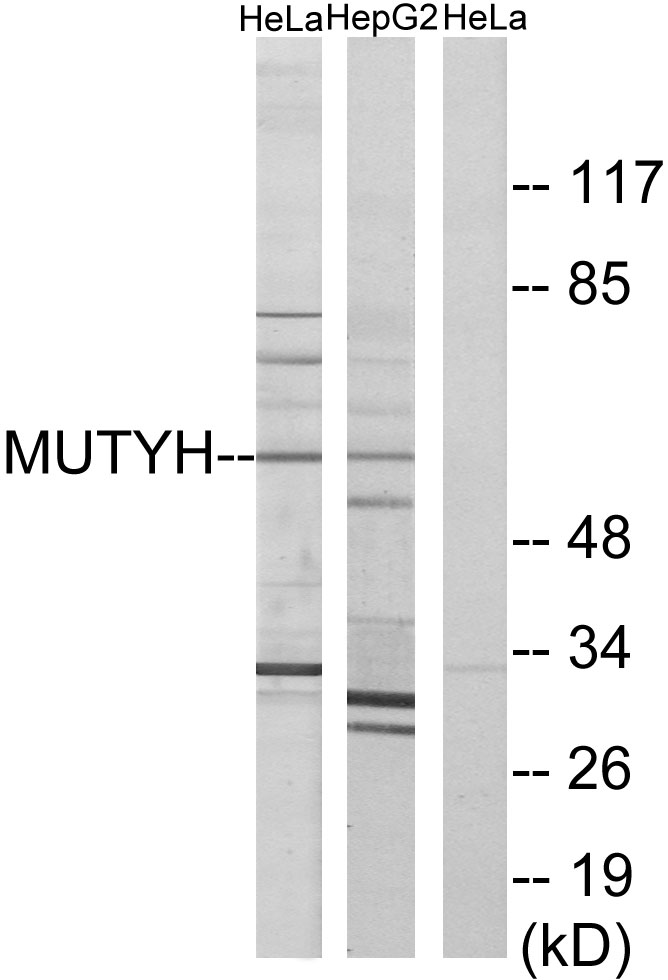
Figure 1. Western blot analysis of MUTYH using anti-MUTYH antibody (A01618-2). Electrophoresis was performed on a 5-20% SDS-PAGE gel at 70V (Stacking gel) / 90V (Resolving gel) for 2-3 hours. The sample well of each lane was loaded with 30 ug of sample under reducing conditions. Lane 1: human Hela whole cell lysates, Lane 2: human K562 whole cell lysates, Lane 3: human HEL whole cell lysates, Lane 4: rat C6 whole cell lysates. After electrophoresis, proteins were transferred to a nitrocellulose membrane at 150 mA for 50-90 minutes. Blocked the membrane with 5% non-fat milk/TBS for 1.5 hour at RT. The membrane was incubated with rabbit anti-MUTYH antigen affinity purified polyclonal antibody (Catalog # A01618-2) at 0.5 microg/mL overnight at 4°C, then washed with TBS-0.1%Tween 3 times with 5 minutes each and probed with a goat anti-rabbit IgG-HRP secondary antibody at a dilution of 1:5000 for 1.5 hour at RT. The signal is developed using an Enhanced Chemiluminescent detection (ECL) kit (Catalog # EK1002) with Tanon 5200 system. A specific band was detected for MUTYH at approximately 60 kDa. The expected band size for MUTYH is at 60 kDa.
Anti-MUTYH Antibody Picoband(r)
A01618-2-CARRIER-FREE
ApplicationsFlow Cytometry, Western Blot, ELISA
Product group Antibodies
ReactivityHuman, Rat
TargetMUTYH
Overview
- SupplierBoster Bio
- Product NameAnti-MUTYH Antibody Picoband(r)
- Delivery Days Customer9
- ApplicationsFlow Cytometry, Western Blot, ELISA
- CertificationResearch Use Only
- ClonalityPolyclonal
- Concentration500 ug/ml
- Gene ID4595
- Target nameMUTYH
- Target descriptionmutY DNA glycosylase
- Target synonymsMYH, adenine DNA glycosylase, A/G-specific adenine DNA glycosylase, mutY homolog, mutY-like protein
- HostRabbit
- IsotypeIgG
- Protein IDQ9UIF7
- Protein NameAdenine DNA glycosylase
- Scientific DescriptionBoster Bio Anti-MUTYH Antibody Picoband® catalog # A01618-2. Tested in ELISA, WB, Flow Cytometry applications. This antibody reacts with Human, Rat. The brand Picoband indicates this is a premium antibody that guarantees superior quality, high affinity, and strong signals with minimal background in Western blot applications. Only our best-performing antibodies are designated as Picoband, ensuring unmatched performance.
- ReactivityHuman, Rat
- Storage Instruction-20°C,2°C to 8°C
- UNSPSC12352203
Related products
Product group Antibodies
Anti-MUTYH AntibodyA96045
ApplicationsImmunoFluorescence, Western Blot, ELISA
ReactivityHuman, Mouse, Rat
- SizePrice
Product group Antibodies
Anti-MUTYH Antibody101-11043
ApplicationsImmunoFluorescence, Western Blot, ELISA
TargetMUTYH
- SizePrice
Product group Antibodies
MUTYH AntibodyCSB-PA003342
ApplicationsImmunoFluorescence, Western Blot, ELISA, ImmunoHistoChemistry
ReactivityHuman, Mouse, Rat
TargetMUTYH
- SizePrice
Product group Antibodies

Goat anti-MUTYHEB06329
ApplicationsWestern Blot, ELISA
ReactivityHuman
TargetMUTYH
- SizePrice
Product group Antibodies
ApplicationsImmunoPrecipitation, Western Blot, ImmunoCytoChemistry, ImmunoHistoChemistry
TargetMUTYH
- SizePrice
Product group Antibodies

MUTYH / MYH AntibodyLS-C400901
ApplicationsWestern Blot, ELISA, ImmunoHistoChemistry
ReactivityHuman
TargetMUTYH
- SizePrice
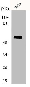
![Various whole cell extracts (30 μg) were separated by 7.5% SDS-PAGE, and the membrane was blotted with MUTYH antibody [N3C3] (GTX103185) diluted at 1:1000. The HRP-conjugated anti-rabbit IgG antibody (GTX213110-01) was used to detect the primary antibody, and the signal was developed with Trident ECL plus-Enhanced.](https://www.genetex.com/upload/website/prouct_img/normal/GTX103185/GTX103185_44643_20220506_WB_w_23060119_265.webp)
Product group Antibodies
MUTYH antibody [N3C3]GTX103185
ApplicationsImmunoFluorescence, Western Blot, ImmunoCytoChemistry
ReactivityHuman, Mouse
TargetMUTYH
- SizePrice

Product group Antibodies
Anti-MUTYH AntibodyHPA008732
ApplicationsWestern Blot, ImmunoCytoChemistry, ImmunoHistoChemistry
ReactivityHuman
TargetMUTYH
- SizePrice
Product group Antibodies
Anti-DNTTIP2 AntibodyCAB16120
ApplicationsImmunoFluorescence, Western Blot, ELISA, ImmunoCytoChemistry
ReactivityHuman
TargetMUTYH
- SizePrice