Bio-Connect
Immunohistochemical staining of human Testis shows moderate nuclear positivity in cells in seminiferous ducts.
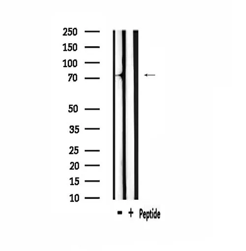
Anti-MYBL1 Antibody

HPA008791
Atlas Antibodies
ApplicationsImmunoHistoChemistry
Product group Antibodies
ReactivityHuman
TargetMYBL1
Sign in to order and to see your custom pricing.
Large volume orders?
Order with a bulk request

Overview

  • Supplier
    Atlas Antibodies
  • Product Name
    Anti-MYBL1
  • Delivery Days Customer
    4
  • Applications
    ImmunoHistoChemistry
  • Certification
    Research Use Only
  • Clonality
    Polyclonal
  • Conjugate
    Unconjugated
  • Gene ID4603
  • Target name
    MYBL1
  • Target description
    MYB proto-oncogene like 1
  • Target synonyms
    AMYB; A-MYB; myb-like protein 1; myb-related protein A; v-myb avian myeloblastosis viral oncogene homolog-like 1
  • Host
    Rabbit
  • Isotype
    IgG
  • Protein IDP10243
  • Protein Name
    Myb-related protein A
  • Scientific Description
    Recombinant Protein Epitope Signature Tag (PrEST) antigen sequence
  • Reactivity
    Human
  • Storage Instruction
    -20°C,2°C to 8°C
  • UNSPSC
    41116161

Related products

Product group Antibodies
Cusabio
MYBL1 AntibodyCSB-PA010296
ApplicationsWestern Blot, ELISA
ReactivityHuman, Mouse
TargetMYBL1
  • SizePrice
Product group Antibodies
LifeSpan BioSciences
ApplicationsWestern Blot
TargetMYBL1
  • SizePrice
Product group Antibodies
Abbexa
ApplicationsWestern Blot, ELISA
  • SizePrice
Product group Antibodies
RayBiotech
ApplicationsWestern Blot
TargetMYBL1
  • SizePrice
Atlas Antibodies
Protein IDP10243
  • SizePrice
ICC/IF analysis of PFA-fixed A549 cells using GTX03441 A-Myb antibody. Permeabilization : 0.1% Triton X-100 Red : Primary antibody Green : beta tubulin Blue : DAPI
Product group Antibodies
GeneTex
ApplicationsImmunoFluorescence, Western Blot, ImmunoCytoChemistry
TargetMYBL1
  • SizePrice
Figure 1. Western blot analysis of v-Myb/MYBL1 using anti-v-Myb/MYBL1 antibody (A05803-2). Electrophoresis was performed on a 5-20% SDS-PAGE gel at 70V (Stacking gel) / 90V (Resolving gel) for 2-3 hours. The sample well of each lane was loaded with 30ug of sample under reducing conditions. Lane 1: human T-47D whole cell lysates, Lane 2: human A431 whole cell lysates, Lane 3: human PC-3 whole cell lysates, Lane 4: human U-87MG whole cell lysates, Lane 5: human A549 whole cell lysates, Lane 6: rat stomach tissue lysates, Lane 7: mouse stomach tissue lysates. After Electrophoresis, proteins were transferred to a Nitrocellulose membrane at 150mA for 50-90 minutes. Blocked the membrane with 5% Non-fat Milk/ TBS for 1.5 hour at RT. The membrane was incubated with rabbit anti-v-Myb/MYBL1 antigen affinity purified polyclonal antibody (Catalog # A05803-2) at 0.5 microg/mL overnight at 4°C, then washed with TBS-0.1%Tween 3 times with 5 minutes each and probed with a goat anti-rabbit IgG-HRP secondary antibody at a dilution of 1:5000 for 1.5 hour at RT. The signal is developed using an Enhanced Chemiluminescent detection (ECL) kit (Catalog # EK1002) with Tanon 5200 system. A specific band was detected for v-Myb/MYBL1 at approximately 86KD. The expected band size for v-Myb/MYBL1 is at 86KD.
Product group Antibodies
Boster Bio
ApplicationsFlow Cytometry, Western Blot, ELISA
TargetMYBL1
  • SizePrice