


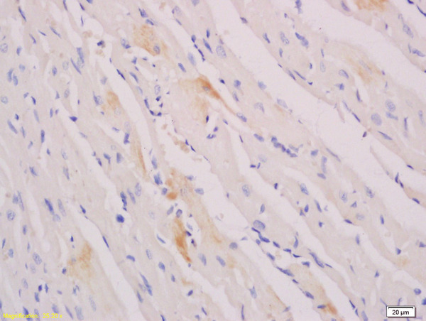
Immunofluorescent staining of human cell line RH-30 shows localization to nucleoplasm.
Immunofluorescent staining of human cell line RH-30 shows localization to nucleoplasm.
Anti-MYOG Antibody
HPA028336
ApplicationsChIP Chromatin ImmunoPrecipitation, ImmunoCytoChemistry
Product group Antibodies
ReactivityHuman
TargetMYOG
Overview
- SupplierAtlas Antibodies
- Product NameAnti-MYOG Antibody
- Delivery Days Customer4
- ApplicationsChIP Chromatin ImmunoPrecipitation, ImmunoCytoChemistry
- CertificationResearch Use Only
- ClonalityPolyclonal
- ConjugateUnconjugated
- Gene ID4656
- Target nameMYOG
- Target descriptionmyogenin
- Target synonymsMYF4, bHLHc3, myf-4, myogenin, class C basic helix-loop-helix protein 3, myogenic factor 4
- HostRabbit
- IsotypeIgG
- Protein IDP15173
- Protein NameMyogenin
- Scientific DescriptionRecombinant Protein Epitope Signature Tag (PrEST) antigen sequence
- ReactivityHuman
- Storage Instruction-20°C,2°C to 8°C
- UNSPSC41116161
Datasheet
MSDS
Related products
Product group Antibodies
MYOG AntibodyCSB-PA003347
ApplicationsWestern Blot, ELISA
ReactivityHuman, Mouse, Rat
TargetMYOG
- SizePrice
Product group Antibodies
Anti-Myogenin AntibodyA96328
ApplicationsWestern Blot, ELISA
ReactivityHuman, Mouse, Rat
- SizePrice

Product group Antibodies
Anti-Myogenin/MYOG Antibody Picoband(r)A03238-1-CARRIER-FREE
ApplicationsWestern Blot, ELISA
ReactivityMonkey, Mouse, Rat
TargetMYOG
- SizePrice
Product group Antibodies

ApplicationsImmunoHistoChemistry
ReactivityHuman
TargetMYOG
- SizePrice
Product group Antibodies

Goat anti-Myogenin / MYF4EB09955
ApplicationsWestern Blot, ELISA
ReactivityBovine, Canine, Human, Mouse, Porcine, Rat
TargetMYOG
- SizePrice
Product group Antibodies
ApplicationsImmunoPrecipitation, Western Blot, ImmunoCytoChemistry, ImmunoHistoChemistry
TargetMYOG
- SizePrice
Product group Antibodies
Myogenin antibodyGTX100887
ApplicationsWestern Blot
ReactivityHuman
TargetMYOG
- SizePrice
Product group Antibodies
References
Myogenin Polyclonal Antibodybs-3550R
ApplicationsImmunoFluorescence, Western Blot, ImmunoCytoChemistry, ImmunoHistoChemistry, ImmunoHistoChemistry Frozen, ImmunoHistoChemistry Paraffin
ReactivityBovine, Human, Mouse, Porcine, Rat
TargetMYOG
- SizePrice