


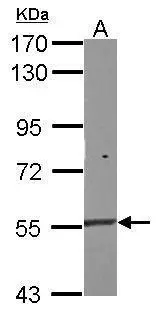
Figure 1. Western blot analysis of Nac1/NACC1 using anti-Nac1/NACC1 antibody (A08675-3). Electrophoresis was performed on a 5-20% SDS-PAGE gel at 70V (Stacking gel) / 90V (Resolving gel) for 2-3 hours. The sample well of each lane was loaded with 30 ug of sample under reducing conditions. Lane 1: human Hela whole cell lysates, Lane 2: human MCF-7 whole cell lysates, Lane 3: human Caco-2 whole cell lysates. After electrophoresis, proteins were transferred to a nitrocellulose membrane at 150 mA for 50-90 minutes. Blocked the membrane with 5% non-fat milk/TBS for 1.5 hour at RT. The membrane was incubated with rabbit anti-Nac1/NACC1 antigen affinity purified polyclonal antibody (Catalog # A08675-3) at 0.5 microg/mL overnight at 4°C, then washed with TBS-0.1%Tween 3 times with 5 minutes each and probed with a goat anti-rabbit IgG-HRP secondary antibody at a dilution of 1:5000 for 1.5 hour at RT. The signal is developed using an Enhanced Chemiluminescent detection (ECL) kit (Catalog # EK1002) with Tanon 5200 system. A specific band was detected for Nac1/NACC1 at approximately 68 kDa. The expected band size for Nac1/NACC1 is at 57 kDa.
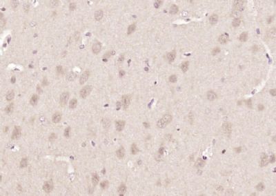
. Nac1/NACC1 was detected in a paraffin-embedded section of human bladder epithelial carcinoma tissue. Heat mediated antigen retrieval was performed in EDTA buffer (pH 8.0, epitope retrieval solution). The tissue section was blocked with 10% goat serum. The tissue section was then incubated with 2 microg/ml rabbit anti-Nac1/NACC1 Antibody (A08675-3) overnight at 4°C. Biotinylated goat anti-rabbit IgG was used as secondary antibody and incubated for 30 minutes at 37°C. The tissue section was developed using Strepavidin-Biotin-Complex (SABC) (Catalog # SA1022) with DAB as the chromogen.")
. Nac1/NACC1 was detected in a paraffin-embedded section of human papillary carcinoma of the left breast tissue. Heat mediated antigen retrieval was performed in EDTA buffer (pH 8.0, epitope retrieval solution). The tissue section was blocked with 10% goat serum. The tissue section was then incubated with 2 microg/ml rabbit anti-Nac1/NACC1 Antibody (A08675-3) overnight at 4°C. Biotinylated goat anti-rabbit IgG was used as secondary antibody and incubated for 30 minutes at 37°C. The tissue section was developed using Strepavidin-Biotin-Complex (SABC) (Catalog # SA1022) with DAB as the chromogen.")
. Nac1/NACC1 was detected in a paraffin-embedded section of human the renal pelvis is squamous metaplasia tissue. Heat mediated antigen retrieval was performed in EDTA buffer (pH 8.0, epitope retrieval solution). The tissue section was blocked with 10% goat serum. The tissue section was then incubated with 2 microg/ml rabbit anti-Nac1/NACC1 Antibody (A08675-3) overnight at 4°C. Biotinylated goat anti-rabbit IgG was used as secondary antibody and incubated for 30 minutes at 37°C. The tissue section was developed using Strepavidin-Biotin-Complex (SABC) (Catalog # SA1022) with DAB as the chromogen.")
. Nac1/NACC1 was detected in an immunocytochemical section of MCF-7 cells. Enzyme antigen retrieval was performed using IHC enzyme antigen retrieval reagent (AR0022) for 15 mins. The cells were blocked with 10% goat serum. And then incubated with 5 microg/mL rabbit anti-Nac1/NACC1 Antibody (A08675-3) overnight at 4°C. DyLight®488 Conjugated Goat Anti-Rabbit IgG (BA1127) was used as secondary antibody at 1:100 dilution and incubated for 30 minutes at 37°C. The tissue section was developed using Phalloidin-iFluor 555 Conjugated. Visualize using a fluorescence microscope and filter sets appropriate for the label used.")
. Overlay histogram showing K562 cells stained with A08675-3 (Blue line). To facilitate intracellular staining, cells were fixed with 4% paraformaldehyde and permeabilized with permeabilization buffer. The cells were blocked with 10% normal goat serum. And then incubated with rabbit anti-Nac1/NACC1 Antibody (A08675-3, 1 microg/1x106 cells) for 30 min at 20°C. DyLight®488 conjugated goat anti-rabbit IgG (BA1127, 5-10 microg/1x106 cells) was used as secondary antibody for 30 minutes at 20°C. Isotype control antibody (Green line) was rabbit IgG (1 microg/1x106) used under the same conditions. Unlabelled sample without incubation with primary antibody and secondary antibody (Red line) was used as a blank control.")
Figure 1. Western blot analysis of Nac1/NACC1 using anti-Nac1/NACC1 antibody (A08675-3). Electrophoresis was performed on a 5-20% SDS-PAGE gel at 70V (Stacking gel) / 90V (Resolving gel) for 2-3 hours. The sample well of each lane was loaded with 30 ug of sample under reducing conditions. Lane 1: human Hela whole cell lysates, Lane 2: human MCF-7 whole cell lysates, Lane 3: human Caco-2 whole cell lysates. After electrophoresis, proteins were transferred to a nitrocellulose membrane at 150 mA for 50-90 minutes. Blocked the membrane with 5% non-fat milk/TBS for 1.5 hour at RT. The membrane was incubated with rabbit anti-Nac1/NACC1 antigen affinity purified polyclonal antibody (Catalog # A08675-3) at 0.5 microg/mL overnight at 4°C, then washed with TBS-0.1%Tween 3 times with 5 minutes each and probed with a goat anti-rabbit IgG-HRP secondary antibody at a dilution of 1:5000 for 1.5 hour at RT. The signal is developed using an Enhanced Chemiluminescent detection (ECL) kit (Catalog # EK1002) with Tanon 5200 system. A specific band was detected for Nac1/NACC1 at approximately 68 kDa. The expected band size for Nac1/NACC1 is at 57 kDa.
Anti-Nac1/NACC1 Antibody Picoband(r)
A08675-3-CARRIER-FREE
ApplicationsFlow Cytometry, ImmunoFluorescence, Western Blot, ELISA, ImmunoCytoChemistry, ImmunoHistoChemistry
Product group Antibodies
ReactivityHuman
TargetNACC1
Overview
- SupplierBoster Bio
- Product NameAnti-Nac1/NACC1 Antibody Picoband(r)
- Delivery Days Customer9
- ApplicationsFlow Cytometry, ImmunoFluorescence, Western Blot, ELISA, ImmunoCytoChemistry, ImmunoHistoChemistry
- CertificationResearch Use Only
- ClonalityPolyclonal
- Concentration500 ug/ml
- Gene ID112939
- Target nameNACC1
- Target descriptionnucleus accumbens associated 1
- Target synonymsBEND8, BTBD14B, BTBD30, NAC-1, NAC1, NECFM, nucleus accumbens-associated protein 1, BEN domain containing 8, BTB/POZ domain-containing protein 14B, nucleus accumbens associated 1, BEN and BTB (POZ) domain containing, transcriptional repressor NAC1
- HostRabbit
- IsotypeIgG
- Protein IDQ96RE7
- Protein NameNucleus accumbens-associated protein 1
- Scientific DescriptionBoster Bio Anti-Nac1/NACC1 Antibody Picoband® catalog # A08675-3. Tested in ELISA, Flow Cytometry, IF, IHC, ICC, WB applications. This antibody reacts with Human. The brand Picoband indicates this is a premium antibody that guarantees superior quality, high affinity, and strong signals with minimal background in Western blot applications. Only our best-performing antibodies are designated as Picoband, ensuring unmatched performance.
- ReactivityHuman
- Storage Instruction-20°C,2°C to 8°C
- UNSPSC12352203
Related products
Product group Antibodies
Anti-NACC1 Antibody144-11720
ApplicationsWestern Blot
ReactivityHuman, Mouse
TargetNACC1
- SizePrice

Product group Antibodies
NAC1 Polyclonal AntibodyBS-12247R
ApplicationsImmunoFluorescence, Western Blot, ELISA, ImmunoCytoChemistry, ImmunoHistoChemistry, ImmunoHistoChemistry Frozen, ImmunoHistoChemistry Paraffin
ReactivityBovine, Canine, Equine, Human, Mouse, Porcine, Rat, Sheep
TargetNACC1
- SizePrice
Product group Antibodies

Goat anti-NAC1 / BTBD14BEB07708
ApplicationsWestern Blot, ELISA
ReactivityCanine, Human
TargetNACC1
- SizePrice
Product group Antibodies
NACC1 AntibodyCSB-PA283286
ApplicationsWestern Blot, ELISA
ReactivityHuman
TargetNACC1
- SizePrice

Product group Antibodies
Anti-NACC1 AntibodyHPA021238
ApplicationsImmunoCytoChemistry, ImmunoHistoChemistry
ReactivityHuman
TargetNACC1
- SizePrice
Product group Antibodies
NAC1 antibodyGTX119455
ApplicationsWestern Blot
ReactivityHuman, Mouse
TargetNACC1
- SizePrice
Product group Antibodies

NACC1 / NAC1 AntibodyLS-C747092
ApplicationsWestern Blot
ReactivityHuman, Mouse, Rat
TargetNACC1
- SizePrice