

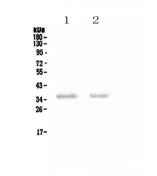
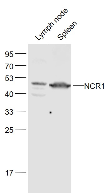
Figure 1. Western blot analysis of NCR1 using anti-NCR1 antibody (A01516-1). Electrophoresis was performed on a 5-20% SDS-PAGE gel at 70V (Stacking gel) / 90V (Resolving gel) for 2-3 hours. The sample well of each lane was loaded with 50ug of sample under reducing conditions. Lane 1: rat spleen tissue lysates, Lane 2: mouse spleen tissue lysates. After Electrophoresis, proteins were transferred to a Nitrocellulose membrane at 150mA for 50-90 minutes. Blocked the membrane with 5% Non-fat Milk/ TBS for 1.5 hour at RT. The membrane was incubated with rabbit anti-NCR1 antigen affinity purified polyclonal antibody (Catalog # A01516-1) at 0.5 microg/mL overnight at 4°C, then washed with TBS-0.1%Tween 3 times with 5 minutes each and probed with a goat anti-rabbit IgG-HRP secondary antibody at a dilution of 1:10000 for 1.5 hour at RT. The signal is developed using an Enhanced Chemiluminescent detection (ECL) kit (Catalog # EK1002) with Tanon 5200 system. A specific band was detected for NCR1 at approximately 36KD. The expected band size for NCR1 is at 34KD.
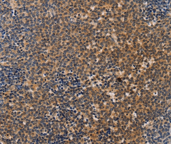
. NCR1 was detected in paraffin-embedded section of rat spleen tissue . Heat mediated antigen retrieval was performed in citrate buffer (pH6, epitope retrieval solution) for 20 mins. The tissue section was blocked with 10% goat serum. The tissue section was then incubated with 1microg/ml rabbit anti-NCR1 Antibody (A01516-1) overnight at 4°C. Biotinylated goat anti-rabbit IgG was used as secondary antibody and incubated for 30 minutes at 37°C. The tissue section was developed using Strepavidin-Biotin-Complex (SABC)(Catalog # SA1022) with DAB as the chromogen.")
. NCR1 was detected in paraffin-embedded section of mouse spleen tissue . Heat mediated antigen retrieval was performed in citrate buffer (pH6, epitope retrieval solution) for 20 mins. The tissue section was blocked with 10% goat serum. The tissue section was then incubated with 1microg/ml rabbit anti-NCR1 Antibody (A01516-1) overnight at 4°C. Biotinylated goat anti-rabbit IgG was used as secondary antibody and incubated for 30 minutes at 37°C. The tissue section was developed using Strepavidin-Biotin-Complex (SABC)(Catalog # SA1022) with DAB as the chromogen.")
. NCR1 was detected in paraffin-embedded section of mouse lung tissues. Heat mediated antigen retrieval was performed in citrate buffer (pH6, epitope retrieval solution) for 20 mins. The tissue section was blocked with 10% goat serum. The tissue section was then incubated with 1microg/ml rabbit anti-NCR1 Antibody (A01516-1) overnight at 4°C. Biotinylated goat anti-rabbit IgG was used as secondary antibody and incubated for 30 minutes at 37°C. The tissue section was developed using Strepavidin-Biotin-Complex (SABC)(Catalog # SA1022) with DAB as the chromogen.")
. NCR1 was detected in paraffin-embedded section of human tonsil tissue. Heat mediated antigen retrieval was performed in EDTA buffer (pH8.0, epitope retrieval solution). The tissue section was blocked with 10% goat serum. The tissue section was then incubated with 1microg/ml rabbit anti-NCR1 Antibody (A01516-1) overnight at 4°C. Biotinylated goat anti-rabbit IgG was used as secondary antibody and incubated for 30 minutes at 37°C. The tissue section was developed using Strepavidin-Biotin-Complex (SABC) (Catalog # SA1022) with DAB as the chromogen.")
Figure 1. Western blot analysis of NCR1 using anti-NCR1 antibody (A01516-1). Electrophoresis was performed on a 5-20% SDS-PAGE gel at 70V (Stacking gel) / 90V (Resolving gel) for 2-3 hours. The sample well of each lane was loaded with 50ug of sample under reducing conditions. Lane 1: rat spleen tissue lysates, Lane 2: mouse spleen tissue lysates. After Electrophoresis, proteins were transferred to a Nitrocellulose membrane at 150mA for 50-90 minutes. Blocked the membrane with 5% Non-fat Milk/ TBS for 1.5 hour at RT. The membrane was incubated with rabbit anti-NCR1 antigen affinity purified polyclonal antibody (Catalog # A01516-1) at 0.5 microg/mL overnight at 4°C, then washed with TBS-0.1%Tween 3 times with 5 minutes each and probed with a goat anti-rabbit IgG-HRP secondary antibody at a dilution of 1:10000 for 1.5 hour at RT. The signal is developed using an Enhanced Chemiluminescent detection (ECL) kit (Catalog # EK1002) with Tanon 5200 system. A specific band was detected for NCR1 at approximately 36KD. The expected band size for NCR1 is at 34KD.
Anti-NCR1 Antibody Picoband(r)
A01516-1-CARRIER-FREE
ApplicationsWestern Blot, ELISA, ImmunoHistoChemistry
Product group Antibodies
ReactivityHuman, Mouse, Rat
TargetNCR1
Overview
- SupplierBoster Bio
- Product NameAnti-NCR1 Antibody Picoband(r)
- Delivery Days Customer9
- ApplicationsWestern Blot, ELISA, ImmunoHistoChemistry
- CertificationResearch Use Only
- ClonalityPolyclonal
- Concentration500 ug/ml
- Gene ID9437
- Target nameNCR1
- Target descriptionnatural cytotoxicity triggering receptor 1
- Target synonymsCD335, LY94, NK-p46, NKP46, natural cytotoxicity triggering receptor 1, NK cell-activating receptor, lymphocyte antigen 94 homolog (activating NK-receptor; NK-p46), natural killer cell p46-related protein
- HostRabbit
- IsotypeIgG
- Protein IDO76036
- Protein NameNatural cytotoxicity triggering receptor 1
- Scientific DescriptionBoster Bio Anti-NCR1 Antibody Picoband® catalog # A01516-1. Tested in ELISA, IHC, WB applications. This antibody reacts with Human, Mouse, Rat. The brand Picoband indicates this is a premium antibody that guarantees superior quality, high affinity, and strong signals with minimal background in Western blot applications. Only our best-performing antibodies are designated as Picoband, ensuring unmatched performance.
- ReactivityHuman, Mouse, Rat
- Storage Instruction-20°C,2°C to 8°C
- UNSPSC12352203
Related products
Product group Antibodies
NCR1 AntibodyCSB-PA003455
ApplicationsWestern Blot, ELISA
ReactivityHuman
TargetNCR1
- SizePrice
Product group Antibodies
Anti-NCR1 AntibodyA101361
ApplicationsWestern Blot, ELISA
ReactivityHuman
- SizePrice
Product group Antibodies
Anti-NCR1 Antibody144-63313
ApplicationsWestern Blot
ReactivityHuman, Mouse, Rat
TargetNCR1
- SizePrice
Product group Antibodies

Anti-NKp46 [Bab281]Ab03165-1.1
ApplicationsFlow Cytometry, ImmunoFluorescence, ImmunoPrecipitation
ReactivityHuman, Primate
TargetNCR1
- SizePrice
Product group Antibodies

ApplicationsFlow Cytometry
ReactivityMouse
TargetNCR1
- SizePrice
Product group Antibodies
NKp46 antibodyGTX05037
ApplicationsWestern Blot, ImmunoHistoChemistry, ImmunoHistoChemistry Paraffin
ReactivityHuman, Mouse
TargetNCR1
- SizePrice
Product group Antibodies
References
NCR1 Polyclonal AntibodyBS-10027R
ApplicationsWestern Blot, ELISA, ImmunoHistoChemistry, ImmunoHistoChemistry Paraffin
ReactivityBovine, Canine, Equine, Human, Mouse, Porcine, Rat, Sheep
TargetNCR1
- SizePrice