

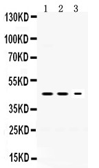
Figure 1. Western blot analysis of Neuroserpin using anti-Neuroserpin antibody (PB9744). Electrophoresis was performed on a 5-20% SDS-PAGE gel at 70V (Stacking gel) / 90V (Resolving gel) for 2-3 hours. Lane 1: Rat Brain Tissue Lysate at 50ug, Lane 2: Mouse Brain Tissue Lysate at 50ug, Lane 3: PANC Whole Cell Lysate at 40ug. After electrophoresis, proteins were transferred to a nitrocellulose membrane at 150 mA for 50-90 minutes. Blocked the membrane with 5% non-fat milk/TBS for 1.5 hour at RT. The membrane was incubated with rabbit anti-Neuroserpin antigen affinity purified polyclonal antibody (Catalog # PB9744) at 0.5 microg/mL overnight at 4°C, then washed with TBS-0.1%Tween 3 times with 5 minutes each and probed with a goat anti-rabbit IgG-HRP secondary antibody at a dilution of 1:5000 for 1.5 hour at RT. The signal is developed using an Enhanced Chemiluminescent detection (ECL) kit (Catalog # EK1002) with Tanon 5200 system. A specific band was detected for Neuroserpin at approximately 46 kDa. The expected band size for Neuroserpin is at 46 kDa.
Figure 1. Western blot analysis of Neuroserpin using anti-Neuroserpin antibody (PB9744). Electrophoresis was performed on a 5-20% SDS-PAGE gel at 70V (Stacking gel) / 90V (Resolving gel) for 2-3 hours. Lane 1: Rat Brain Tissue Lysate at 50ug, Lane 2: Mouse Brain Tissue Lysate at 50ug, Lane 3: PANC Whole Cell Lysate at 40ug. After electrophoresis, proteins were transferred to a nitrocellulose membrane at 150 mA for 50-90 minutes. Blocked the membrane with 5% non-fat milk/TBS for 1.5 hour at RT. The membrane was incubated with rabbit anti-Neuroserpin antigen affinity purified polyclonal antibody (Catalog # PB9744) at 0.5 microg/mL overnight at 4°C, then washed with TBS-0.1%Tween 3 times with 5 minutes each and probed with a goat anti-rabbit IgG-HRP secondary antibody at a dilution of 1:5000 for 1.5 hour at RT. The signal is developed using an Enhanced Chemiluminescent detection (ECL) kit (Catalog # EK1002) with Tanon 5200 system. A specific band was detected for Neuroserpin at approximately 46 kDa. The expected band size for Neuroserpin is at 46 kDa.
Anti-Neuroserpin/SERPINI1 Antibody Picoband(r)
PB9744-CARRIER-FREE
ApplicationsWestern Blot
Product group Antibodies
ReactivityHuman, Mouse, Rat
TargetSERPINI1
Overview
- SupplierBoster Bio
- Product NameAnti-Neuroserpin/SERPINI1 Antibody Picoband(r)
- Delivery Days Customer9
- Application Supplier NoteTested Species: In-house tested species with positive results. Other applications have not been tested. Optimal dilutions should be determined by end users.
- ApplicationsWestern Blot
- CertificationResearch Use Only
- ClonalityPolyclonal
- Concentration500 ug/ml
- Gene ID5274
- Target nameSERPINI1
- Target descriptionserpin family I member 1
- Target synonymsHNS-S1, HNS-S2, PI12, neuroserpin, neuroserpin, PI-12, peptidase inhibitor 12, serine (or cysteine) proteinase inhibitor, clade I (neuroserpin), member 1, serpin I1, serpin peptidase inhibitor clade I member 1, serpin peptidase inhibitor, clade I (neuroserpin), member 1
- HostRabbit
- IsotypeIgG
- Protein IDQ99574
- Protein NameNeuroserpin
- Scientific DescriptionBoster Bio Anti-Neuroserpin/SERPINI1 Antibody Picoband® catalog # PB9744. Tested in WB applications. This antibody reacts with Human, Mouse, Rat. The brand Picoband indicates this is a premium antibody that guarantees superior quality, high affinity, and strong signals with minimal background in Western blot applications. Only our best-performing antibodies are designated as Picoband, ensuring unmatched performance.
- ReactivityHuman, Mouse, Rat
- Storage Instruction-20°C,2°C to 8°C
- UNSPSC12352203
Related products
Product group Antibodies
Anti-SERPINI1 Antibody144-61157
ApplicationsWestern Blot, ImmunoHistoChemistry
ReactivityHuman, Mouse, Rat
TargetSERPINI1
- SizePrice
Product group Antibodies
ApplicationsWestern Blot
ReactivityHuman, Mouse, Rat
- SizePrice
Product group Antibodies

Neuroserpin Antibody (aa181-410)LS-C783535
ApplicationsImmunoFluorescence, Western Blot, ImmunoHistoChemistry
ReactivityMouse, Rat
TargetSERPINI1
- SizePrice
Product group Antibodies
Neuroserpin Polyclonal AntibodyBS-11317R
ApplicationsImmunoFluorescence, Western Blot, ELISA, ImmunoCytoChemistry, ImmunoHistoChemistry, ImmunoHistoChemistry Frozen, ImmunoHistoChemistry Paraffin
ReactivityBovine, Canine, Chicken, Equine, Human, Mouse, Porcine, Rabbit, Rat, Sheep
TargetSERPINI1
- SizePrice
Product group Antibodies
SERPINI1 AntibodyCSB-PA859934LA01HU
ApplicationsELISA, ImmunoHistoChemistry
ReactivityHuman
TargetSERPINI1
- SizePrice
Product group Antibodies

Goat anti-NeuroserpinEB06688
ApplicationsFlow Cytometry, Western Blot, ELISA, ImmunoHistoChemistry
ReactivityBovine, Human, Mouse, Porcine, Rat
TargetSERPINI1
- SizePrice
Product group Antibodies
ApplicationsImmunoPrecipitation, Western Blot, ImmunoCytoChemistry, ImmunoHistoChemistry
ReactivityMouse, Porcine, Rat
TargetSERPINI1
- SizePrice
Product group Antibodies
Neuroserpin antibody, C-termGTX89544
ApplicationsWestern Blot, ImmunoHistoChemistry, ImmunoHistoChemistry Paraffin
ReactivityHuman
TargetSERPINI1
- SizePrice

Product group Antibodies
Anti-SERPINI1 AntibodyHPA056092
ApplicationsImmunoHistoChemistry
ReactivityHuman
TargetSERPINI1
- SizePrice