


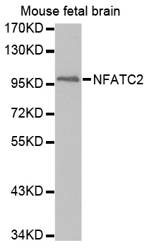
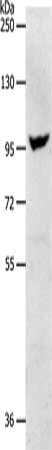
Figure 1. Western blot analysis of NFAT1 using anti-NFAT1 antibody (A00969). Electrophoresis was performed on a 5-20% SDS-PAGE gel at 70V (Stacking gel) / 90V (Resolving gel) for 2-3 hours. The sample well of each lane was loaded with 50ug of sample under reducing conditions. Lane 1: human K562 whole cell lysates. After Electrophoresis, proteins were transferred to a Nitrocellulose membrane at 150mA for 50-90 minutes. Blocked the membrane with 5% Non-fat Milk/ TBS for 1.5 hour at RT. The membrane was incubated with rabbit anti-NFAT1 antigen affinity purified polyclonal antibody (Catalog # A00969) at 0.5 ug/mL overnight at 4 then washed with TBS-0.1%Tween 3 times with 5 minutes each and probed with a goat anti-rabbit IgG-HRP secondary antibody at a dilution of 1:10000 for 1.5 hour at RT. The signal is developed using an Enhanced Chemiluminescent detection (ECL) kit (Catalog # EK1002) with Tanon 5200 system. A specific band was detected for NFAT1 at approximately 100KD. The expected band size for NFAT1 is at 100KD.
Figure 1. Western blot analysis of NFAT1 using anti-NFAT1 antibody (A00969). Electrophoresis was performed on a 5-20% SDS-PAGE gel at 70V (Stacking gel) / 90V (Resolving gel) for 2-3 hours. The sample well of each lane was loaded with 50ug of sample under reducing conditions. Lane 1: human K562 whole cell lysates. After Electrophoresis, proteins were transferred to a Nitrocellulose membrane at 150mA for 50-90 minutes. Blocked the membrane with 5% Non-fat Milk/ TBS for 1.5 hour at RT. The membrane was incubated with rabbit anti-NFAT1 antigen affinity purified polyclonal antibody (Catalog # A00969) at 0.5 ug/mL overnight at 4 then washed with TBS-0.1%Tween 3 times with 5 minutes each and probed with a goat anti-rabbit IgG-HRP secondary antibody at a dilution of 1:10000 for 1.5 hour at RT. The signal is developed using an Enhanced Chemiluminescent detection (ECL) kit (Catalog # EK1002) with Tanon 5200 system. A specific band was detected for NFAT1 at approximately 100KD. The expected band size for NFAT1 is at 100KD.
Anti-NFAT1/NFATC2 Antibody Picoband(r)
A00969-CARRIER-FREE
ApplicationsFlow Cytometry, ImmunoFluorescence, Western Blot, ELISA, ImmunoCytoChemistry
Product group Antibodies
ReactivityHuman
TargetNFATC2
Overview
- SupplierBoster Bio
- Product NameAnti-NFAT1/NFATC2 Antibody Picoband(r)
- Delivery Days Customer9
- Application Supplier NoteTested Species: In-house tested species with positive results. Other applications have not been tested. Optimal dilutions should be determined by end users.
- ApplicationsFlow Cytometry, ImmunoFluorescence, Western Blot, ELISA, ImmunoCytoChemistry
- CertificationResearch Use Only
- ClonalityPolyclonal
- Concentration500 ug/ml
- Gene ID4773
- Target nameNFATC2
- Target descriptionnuclear factor of activated T cells 2
- Target synonymsJCOSL, NFAT1, NFATP, nuclear factor of activated T-cells, cytoplasmic 2, NF-ATc2, NFAT pre-existing subunit, NFAT transcription complex, preexisting component, T cell transcription factor NFAT1, nuclear factor of activated T-cells, cytoplasmic, calcineurin-dependent 2, nuclear factor of activated T-cells, preexisting component, preexisting nuclear factor of activated T-cells 2
- HostRabbit
- IsotypeIgG
- Protein IDQ13469
- Protein NameNuclear factor of activated T-cells, cytoplasmic 2
- Scientific DescriptionBoster Bio Anti-NFAT1/NFATC2 Antibody Picoband® catalog # A00969. Tested in ELISA, Flow Cytometry, IF, ICC, WB applications. This antibody reacts with Human. The brand Picoband indicates this is a premium antibody that guarantees superior quality, high affinity, and strong signals with minimal background in Western blot applications. Only our best-performing antibodies are designated as Picoband, ensuring unmatched performance.
- ReactivityHuman
- Storage Instruction-20°C,2°C to 8°C
- UNSPSC12352203
Related products
Product group Antibodies
Anti-NFATC2 AntibodyA30696
ApplicationsWestern Blot
ReactivityHuman, Mouse, Rat
- SizePrice
Product group Antibodies
anti-NFATc2 (human), pAb (IG-209)AG-25T-0111
ApplicationsImmunoPrecipitation, Western Blot, ImmunoCytoChemistry, ImmunoHistoChemistry
ReactivityHuman
TargetNFATC2
- SizePrice
Product group Antibodies
Anti-NFATC2 Antibody144-03107
ApplicationsWestern Blot
ReactivityHuman, Mouse
TargetNFATC2
- SizePrice
Product group Antibodies
NFATc2 Recombinant Antibody, AbBy Fluor-350 ConjugatedBSM-61447R-BF350
ApplicationsWestern Blot
ReactivityHuman
TargetNFATC2
- SizePrice
Product group Antibodies
NFATC2 AntibodyCSB-PA108395
ApplicationsWestern Blot, ELISA
ReactivityHuman, Mouse
TargetNFATC2
- SizePrice
Product group Antibodies

Goat anti-NFATC2 / NFAT1EB09503
ApplicationsWestern Blot, ELISA, ImmunoHistoChemistry
ReactivityBovine, Canine, Human, Mouse, Rat
TargetNFATC2
- SizePrice
Product group Antibodies
ApplicationsImmunoPrecipitation, Western Blot, ImmunoCytoChemistry, ImmunoHistoChemistry
ReactivityMouse, Rat
TargetNFATC2
- SizePrice
Product group Antibodies

NFAT1 / NFATC2 AntibodyLS-C402704
ApplicationsWestern Blot, ELISA
ReactivityHuman, Mouse
TargetNFATC2
- SizePrice

Product group Antibodies
Anti-NFATC2 AntibodyHPA008789
ApplicationsWestern Blot, ImmunoCytoChemistry, ImmunoHistoChemistry
ReactivityHuman
TargetNFATC2
- SizePrice
Product group Antibodies
NFAT1 antibodyGTX33354
ApplicationsImmunoFluorescence, Western Blot, ImmunoCytoChemistry, ImmunoHistoChemistry, ImmunoHistoChemistry Paraffin
ReactivityHuman, Mouse, Rat
TargetNFATC2
- SizePrice