

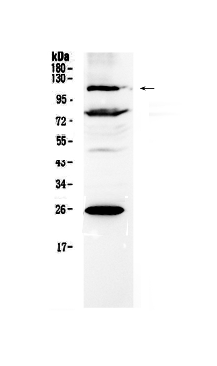
Figure 1. Western blot analysis of NFAT4 using anti-NFAT4 antibody (A02727-2). Electrophoresis was performed on a 5-20% SDS-PAGE gel at 70V (Stacking gel) / 90V (Resolving gel) for 2-3 hours. The sample well of each lane was loaded with 50ug of sample under reducing conditions. Lane 1: human 22RV1 whole cell lysates. After Electrophoresis, proteins were transferred to a Nitrocellulose membrane at 150mA for 50-90 minutes. Blocked the membrane with 5% Non-fat Milk/ TBS for 1.5 hour at RT. The membrane was incubated with rabbit anti-NFAT4 antigen affinity purified polyclonal antibody (Catalog # A02727-2) at 0.5 ug/mL overnight at 4 then washed with TBS-0.1%Tween 3 times with 5 minutes each and probed with a goat anti-rabbit IgG-HRP secondary antibody at a dilution of 1:10000 for 1.5 hour at RT. The signal is developed using an Enhanced Chemiluminescent detection (ECL) kit (Catalog # EK1002) with Tanon 5200 system. A specific band was detected for NFAT4 at approximately 115KD. The expected band size for NFAT4 is at 115KD.
. Overlay histogram showing A431 cells stained with A02727-2 (Blue line). To facilitate intracellular staining, cells were fixed with 4% paraformaldehyde and permeabilized with permeabilization buffer. The cells were blocked with 10% normal goat serum. And then incubated with rabbit anti-NFAT4 Antibody (A02727-2,1microg/1x106 cells) for 30 min at 20°C. DyLight®488 conjugated goat anti-rabbit IgG (BA1127, 5-10microg/1x106 cells) was used as secondary antibody for 30 minutes at 20°C. Isotype control antibody (Green line) was rabbit IgG (1microg/1x106) used under the same conditions. Unlabelled sample without incubation with primary antibody and secondary antibody (Red line) was used as a blank control.")
. Overlay histogram showing U20S cells stained with A02727-2 (Blue line). To facilitate intracellular staining, cells were fixed with 4% paraformaldehyde and permeabilized with permeabilization buffer. The cells were blocked with 10% normal goat serum. And then incubated with rabbit anti-NFAT4 Antibody (A02727-2,1microg/1x106 cells) for 30 min at 20°C. DyLight®488 conjugated goat anti-rabbit IgG (BA1127, 5-10microg/1x106 cells) was used as secondary antibody for 30 minutes at 20°C. Isotype control antibody (Green line) was rabbit IgG (1microg/1x106) used under the same conditions. Unlabelled sample without incubation with primary antibody and secondary antibody (Red line) was used as a blank control.")
. Overlay histogram showing K562 cells stained with A02727-2 (Blue line). To facilitate intracellular staining, cells were fixed with 4% paraformaldehyde and permeabilized with permeabilization buffer. The cells were blocked with 10% normal goat serum. And then incubated with rabbit anti-NFAT4 Antibody (A02727-2,1microg/1x106 cells) for 30 min at 20°C. DyLight®488 conjugated goat anti-rabbit IgG (BA1127, 5-10microg/1x106 cells) was used as secondary antibody for 30 minutes at 20°C. Isotype control antibody (Green line) was rabbit IgG (1microg/1x106) used under the same conditions. Unlabelled sample without incubation with primary antibody and secondary antibody (Red line) was used as a blank control.")
. Overlay histogram showing SiHa cells stained with A02727-2 (Blue line). To facilitate intracellular staining, cells were fixed with 4% paraformaldehyde and permeabilized with permeabilization buffer. The cells were blocked with 10% normal goat serum. And then incubated with rabbit anti-NFAT4 Antibody (A02727-2,1microg/1x106 cells) for 30 min at 20°C. DyLight®488 conjugated goat anti-rabbit IgG (BA1127, 5-10microg/1x106 cells) was used as secondary antibody for 30 minutes at 20°C. Isotype control antibody (Green line) was rabbit IgG (1microg/1x106) used under the same conditions. Unlabelled sample without incubation with primary antibody and secondary antibody (Red line) was used as a blank control.")
Figure 1. Western blot analysis of NFAT4 using anti-NFAT4 antibody (A02727-2). Electrophoresis was performed on a 5-20% SDS-PAGE gel at 70V (Stacking gel) / 90V (Resolving gel) for 2-3 hours. The sample well of each lane was loaded with 50ug of sample under reducing conditions. Lane 1: human 22RV1 whole cell lysates. After Electrophoresis, proteins were transferred to a Nitrocellulose membrane at 150mA for 50-90 minutes. Blocked the membrane with 5% Non-fat Milk/ TBS for 1.5 hour at RT. The membrane was incubated with rabbit anti-NFAT4 antigen affinity purified polyclonal antibody (Catalog # A02727-2) at 0.5 ug/mL overnight at 4 then washed with TBS-0.1%Tween 3 times with 5 minutes each and probed with a goat anti-rabbit IgG-HRP secondary antibody at a dilution of 1:10000 for 1.5 hour at RT. The signal is developed using an Enhanced Chemiluminescent detection (ECL) kit (Catalog # EK1002) with Tanon 5200 system. A specific band was detected for NFAT4 at approximately 115KD. The expected band size for NFAT4 is at 115KD.
Anti-NFAT4/NFATC3 Antibody Picoband(r)
A02727-2-CARRIER-FREE
ApplicationsFlow Cytometry, Western Blot, ELISA, ImmunoCytoChemistry, ImmunoHistoChemistry
Product group Antibodies
ReactivityHuman, Mouse, Rat
TargetNFATC3
Overview
- SupplierBoster Bio
- Product NameAnti-NFAT4/NFATC3 Antibody Picoband(r)
- Delivery Days Customer9
- ApplicationsFlow Cytometry, Western Blot, ELISA, ImmunoCytoChemistry, ImmunoHistoChemistry
- CertificationResearch Use Only
- ClonalityPolyclonal
- Concentration500 ug/ml
- Gene ID4775
- Target nameNFATC3
- Target descriptionnuclear factor of activated T cells 3
- Target synonymsNF-AT4c, NFAT4, NFATX, n339260, nuclear factor of activated T-cells, cytoplasmic 3, NF-ATc3, T cell transcription factor NFAT4, nuclear factor of activated T-cells, cytoplasmic, calcineurin-dependent 3
- HostRabbit
- IsotypeIgG
- Protein IDQ12968
- Protein NameNuclear factor of activated T-cells, cytoplasmic 3
- Scientific DescriptionBoster Bio Anti-NFAT4/NFATC3 Antibody Picoband® catalog # A02727-2. Tested in ELISA, Flow Cytometry, IHC, ICC, WB applications. This antibody reacts with Human, Mouse, Rat. The brand Picoband indicates this is a premium antibody that guarantees superior quality, high affinity, and strong signals with minimal background in Western blot applications. Only our best-performing antibodies are designated as Picoband, ensuring unmatched performance.
- ReactivityHuman, Mouse, Rat
- Storage Instruction-20°C,2°C to 8°C
- UNSPSC12352203
Related products
Product group Antibodies
Anti-NFATC3 AntibodyA39602
ApplicationsImmunoFluorescence, Western Blot
ReactivityHuman, Mouse
- SizePrice
Product group Antibodies
Anti-NFATC3 Antibody144-06666
ApplicationsImmunoFluorescence, Western Blot
ReactivityHuman, Mouse
TargetNFATC3
- SizePrice

Product group Antibodies
ApplicationsImmunoFluorescence, Western Blot, ELISA, ImmunoCytoChemistry, ImmunoHistoChemistry, ImmunoHistoChemistry Frozen, ImmunoHistoChemistry Paraffin
ReactivityBovine, Chicken, Equine, Human, Mouse, Rabbit, Sheep
TargetNFATC3
- SizePrice
Product group Antibodies
NFATC3 AntibodyCSB-PA003421
ApplicationsWestern Blot, ELISA, ImmunoHistoChemistry
ReactivityHuman, Mouse
TargetNFATC3
- SizePrice
Product group Antibodies

Goat anti-NFATC3 / NFAT4EB09504
ApplicationsWestern Blot, ELISA
ReactivityBovine, Canine, Human
TargetNFATC3
- SizePrice
Product group Antibodies
Nfatc3 Polyclonal AntibodyCAC11250
ApplicationsImmunoFluorescence, Western Blot, ELISA, ImmunoHistoChemistry
ReactivityRat
TargetNFATC3
- SizePrice
Product group Antibodies

NFAT4 / NFATC3 AntibodyLS-C400911
ApplicationsELISA, ImmunoHistoChemistry
ReactivityHuman, Mouse
TargetNFATC3
- SizePrice

Product group Antibodies
Anti-NFATC3 AntibodyHPA050665
ApplicationsImmunoCytoChemistry
ReactivityHuman
TargetNFATC3
- SizePrice
Product group Antibodies
NFAT4 (phospho Ser165) antibodyGTX52339
ApplicationsImmunoFluorescence, Western Blot, ImmunoCytoChemistry
ReactivityHuman, Mouse
TargetNFATC3
- SizePrice