


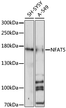
Figure 1. Western blot analysis of NFAT5 using anti-NFAT5 antibody (A01815-3). Electrophoresis was performed on a 5-20% SDS-PAGE gel at 70V (Stacking gel) / 90V (Resolving gel) for 2-3 hours. The sample well of each lane was loaded with 30ug of sample under reducing conditions. Lane 1: human THP-1 whole cell lysates. After Electrophoresis, proteins were transferred to a Nitrocellulose membrane at 150mA for 50-90 minutes. Blocked the membrane with 5% Non-fat Milk/ TBS for 1.5 hour at RT. The membrane was incubated with rabbit anti-NFAT5 antigen affinity purified polyclonal antibody (Catalog # A01815-3) at 0.5 microg/mL overnight at 4°C, then washed with TBS-0.1%Tween 3 times with 5 minutes each and probed with a goat anti-rabbit IgG-HRP secondary antibody at a dilution of 1:5000 for 1.5 hour at RT. The signal is developed using an Enhanced Chemiluminescent detection (ECL) kit (Catalog # EK1002) with Tanon 5200 system. A specific band was detected for NFAT5 at approximately 170KD. The expected band size for NFAT5 is at 170KD.
. Overlay histogram showing HepG2 cells stained with A01815-3 (Blue line). To facilitate intracellular staining, cells were fixed with 4% paraformaldehyde and permeabilized with permeabilization buffer. The cells were blocked with 10% normal goat serum. And then incubated with rabbit anti-NFAT5 Antibody (A01815-3, 1microg/1x106 cells) for 30 min at 20°C. DyLight®488 conjugated goat anti-rabbit IgG (BA1127, 5-10microg/1x106 cells) was used as secondary antibody for 30 minutes at 20°C. Isotype control antibody (Green line) was rabbit IgG (1microg/1x106) used under the same conditions. Unlabelled sample without incubation with primary antibody and secondary antibody (Red line) was used as a blank control.")
Figure 1. Western blot analysis of NFAT5 using anti-NFAT5 antibody (A01815-3). Electrophoresis was performed on a 5-20% SDS-PAGE gel at 70V (Stacking gel) / 90V (Resolving gel) for 2-3 hours. The sample well of each lane was loaded with 30ug of sample under reducing conditions. Lane 1: human THP-1 whole cell lysates. After Electrophoresis, proteins were transferred to a Nitrocellulose membrane at 150mA for 50-90 minutes. Blocked the membrane with 5% Non-fat Milk/ TBS for 1.5 hour at RT. The membrane was incubated with rabbit anti-NFAT5 antigen affinity purified polyclonal antibody (Catalog # A01815-3) at 0.5 microg/mL overnight at 4°C, then washed with TBS-0.1%Tween 3 times with 5 minutes each and probed with a goat anti-rabbit IgG-HRP secondary antibody at a dilution of 1:5000 for 1.5 hour at RT. The signal is developed using an Enhanced Chemiluminescent detection (ECL) kit (Catalog # EK1002) with Tanon 5200 system. A specific band was detected for NFAT5 at approximately 170KD. The expected band size for NFAT5 is at 170KD.
Anti-NFAT5 Antibody Picoband(r)
A01815-3-CARRIER-FREE
ApplicationsFlow Cytometry, ImmunoFluorescence, Western Blot, ELISA, ImmunoCytoChemistry, ImmunoHistoChemistry
Product group Antibodies
ReactivityHuman, Mouse, Rat
TargetNFAT5
Overview
- SupplierBoster Bio
- Product NameAnti-NFAT5 Antibody Picoband(r)
- Delivery Days Customer9
- ApplicationsFlow Cytometry, ImmunoFluorescence, Western Blot, ELISA, ImmunoCytoChemistry, ImmunoHistoChemistry
- CertificationResearch Use Only
- ClonalityPolyclonal
- Concentration500 ug/ml
- Gene ID10725
- Target nameNFAT5
- Target descriptionnuclear factor of activated T cells 5
- Target synonymsNF-AT5, NFATL1, NFATZ, OREBP, TONEBP, nuclear factor of activated T-cells 5, NFAT-like protein 1, T-cell transcription factor NFAT5, TonE-binding protein, glutamine rich protein H65, nuclear factor of activated T-cells 5, tonicity-responsive, osmotic response element-binding protein, tonicity-responsive enhancer-binding protein
- HostRabbit
- IsotypeIgG
- Protein IDO94916
- Protein NameNuclear factor of activated T-cells 5
- Scientific DescriptionBoster Bio Anti-NFAT5 Antibody Picoband® catalog # A01815-3. Tested in ELISA, Flow Cytometry, ICC, IHC, IF, WB applications. This antibody reacts with Human, Mouse, Rat. The brand Picoband indicates this is a premium antibody that guarantees superior quality, high affinity, and strong signals with minimal background in Western blot applications. Only our best-performing antibodies are designated as Picoband, ensuring unmatched performance.
- ReactivityHuman, Mouse, Rat
- Storage Instruction-20°C,2°C to 8°C
- UNSPSC12352203
Related products
Product group Antibodies
Phospho-NFAT5 (S155) AntibodyCSB-PA006686
ApplicationsWestern Blot, ELISA
ReactivityHuman, Mouse
TargetNFAT5
- SizePrice
Product group Antibodies
Anti-NFAT5 Antibody144-64565
ApplicationsWestern Blot
ReactivityHuman
TargetNFAT5
- SizePrice

Product group Antibodies
Anti-NFAT5 AntibodyHPA069711
ApplicationsChIP Chromatin ImmunoPrecipitation, ImmunoCytoChemistry, ImmunoHistoChemistry
ReactivityHuman
TargetNFAT5
- SizePrice
Product group Antibodies

NFAT5 AntibodyLS-C678808
ApplicationsImmunoFluorescence, ELISA
ReactivityHuman
TargetNFAT5
- SizePrice
Product group Antibodies
NFAT5 antibody [N1N2], N-termGTX110903
ApplicationsWestern Blot
ReactivityHuman
TargetNFAT5
- SizePrice
Product group Antibodies
References
NFAT5 Polyclonal AntibodyBS-9473R
ApplicationsImmunoFluorescence, Western Blot, ELISA, ImmunoCytoChemistry, ImmunoHistoChemistry, ImmunoHistoChemistry Frozen, ImmunoHistoChemistry Paraffin
ReactivityHuman, Mouse, Rat
TargetNFAT5
- SizePrice