


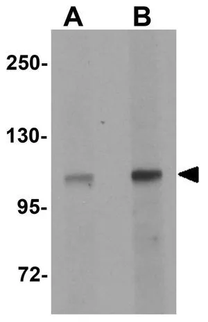
Figure 1. Western blot analysis of NLRX1 using anti-NLRX1 antibody (A04980-3). Electrophoresis was performed on a 5-20% SDS-PAGE gel at 70V (Stacking gel) / 90V (Resolving gel) for 2-3 hours. The sample well of each lane was loaded with 30 ug of sample under reducing conditions. Lane 1: human 293T whole cell lysates, Lane 2: human HepG2 whole cell lysates, Lane 3: human MCF-7 whole cell lysates, Lane 4: human Hela whole cell lysates, Lane 5: rat heart tissue lysates, Lane 6: rat skeletal muscle tissue lysates, Lane 7: mouse heart tissue lysates, Lane 8: mouse skeletal muscle tissue lysates. After electrophoresis, proteins were transferred to a nitrocellulose membrane at 150 mA for 50-90 minutes. Blocked the membrane with 5% non-fat milk/TBS for 1.5 hour at RT. The membrane was incubated with rabbit anti-NLRX1 antigen affinity purified polyclonal antibody (Catalog # A04980-3) at 0.5 microg/mL overnight at 4°C, then washed with TBS-0.1%Tween 3 times with 5 minutes each and probed with a goat anti-rabbit IgG-HRP secondary antibody at a dilution of 1:5000 for 1.5 hour at RT. The signal is developed using an Enhanced Chemiluminescent detection (ECL) kit (Catalog # EK1002) with Tanon 5200 system. A specific band was detected for NLRX1 at approximately 108 kDa. The expected band size for NLRX1 is at 108 kDa.
. NLRX1 was detected in a paraffin-embedded section of human breast cancer tissue. Heat mediated antigen retrieval was performed in EDTA buffer (pH 8.0, epitope retrieval solution). The tissue section was blocked with 10% goat serum. The tissue section was then incubated with 2 microg/ml rabbit anti-NLRX1 Antibody (A04980-3) overnight at 4°C. Peroxidase Conjugated Goat Anti-rabbit IgG was used as secondary antibody and incubated for 30 minutes at 37°C. The tissue section was developed using HRP Conjugated Rabbit IgG Super Vision Assay Kit (Catalog # SV0002) with DAB as the chromogen.")
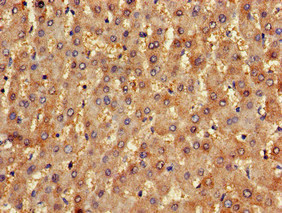
. NLRX1 was detected in a paraffin-embedded section of human lung adenocarcinoma tissue. Heat mediated antigen retrieval was performed in EDTA buffer (pH 8.0, epitope retrieval solution). The tissue section was blocked with 10% goat serum. The tissue section was then incubated with 2 microg/ml rabbit anti-NLRX1 Antibody (A04980-3) overnight at 4°C. Peroxidase Conjugated Goat Anti-rabbit IgG was used as secondary antibody and incubated for 30 minutes at 37°C. The tissue section was developed using HRP Conjugated Rabbit IgG Super Vision Assay Kit (Catalog # SV0002) with DAB as the chromogen.")
. NLRX1 was detected in a paraffin-embedded section of human thyroid papillary carcinoma tissue. Heat mediated antigen retrieval was performed in EDTA buffer (pH 8.0, epitope retrieval solution). The tissue section was blocked with 10% goat serum. The tissue section was then incubated with 2 microg/ml rabbit anti-NLRX1 Antibody (A04980-3) overnight at 4°C. Peroxidase Conjugated Goat Anti-rabbit IgG was used as secondary antibody and incubated for 30 minutes at 37°C. The tissue section was developed using HRP Conjugated Rabbit IgG Super Vision Assay Kit (Catalog # SV0002) with DAB as the chromogen.")
. NLRX1 was detected in an immunocytochemical section of Hela cells. Enzyme antigen retrieval was performed using IHC enzyme antigen retrieval reagent (AR0022) for 15 mins. The cells were blocked with 10% goat serum. And then incubated with 5 microg/mL rabbit anti-NLRX1 Antibody (A04980-3) overnight at 4°C. Cy3 Conjugated Goat Anti-Rabbit IgG (BA1032) was used as secondary antibody at 1:500 dilution and incubated for 30 minutes at 37°C. The section was counterstained with DAPI. Visualize using a fluorescence microscope and filter sets appropriate for the label used.")
. NLRX1 was detected in a paraffin-embedded section of human intestinal cancer tissue. Heat mediated antigen retrieval was performed in EDTA buffer (pH 8.0, epitope retrieval solution). The tissue section was blocked with 10% goat serum. The tissue section was then incubated with 5 microg/mL rabbit anti-NLRX1 Antibody (A04980-3) overnight at 4°C. Cy3 Conjugated Goat Anti-Rabbit IgG (BA1032) was used as secondary antibody at 1:500 dilution and incubated for 30 minutes at 37°C. The section was counterstained with DAPI. Visualize using a fluorescence microscope and filter sets appropriate for the label used.")
. Overlay histogram showing HL-60 cells stained with A04980-3 (Blue line). To facilitate intracellular staining, cells were fixed with 4% paraformaldehyde and permeabilized with permeabilization buffer. The cells were blocked with 10% normal goat serum. And then incubated with rabbit anti-NLRX1 Antibody (A04980-3, 1 microg/1x106 cells) for 30 min at 20°C. DyLight®488 conjugated goat anti-rabbit IgG (BA1127, 5-10 microg/1x106 cells) was used as secondary antibody for 30 minutes at 20°C. Isotype control antibody (Green line) was rabbit IgG (1 microg/1x106) used under the same conditions. Unlabelled sample without incubation with primary antibody and secondary antibody (Red line) was used as a blank control.")
Figure 1. Western blot analysis of NLRX1 using anti-NLRX1 antibody (A04980-3). Electrophoresis was performed on a 5-20% SDS-PAGE gel at 70V (Stacking gel) / 90V (Resolving gel) for 2-3 hours. The sample well of each lane was loaded with 30 ug of sample under reducing conditions. Lane 1: human 293T whole cell lysates, Lane 2: human HepG2 whole cell lysates, Lane 3: human MCF-7 whole cell lysates, Lane 4: human Hela whole cell lysates, Lane 5: rat heart tissue lysates, Lane 6: rat skeletal muscle tissue lysates, Lane 7: mouse heart tissue lysates, Lane 8: mouse skeletal muscle tissue lysates. After electrophoresis, proteins were transferred to a nitrocellulose membrane at 150 mA for 50-90 minutes. Blocked the membrane with 5% non-fat milk/TBS for 1.5 hour at RT. The membrane was incubated with rabbit anti-NLRX1 antigen affinity purified polyclonal antibody (Catalog # A04980-3) at 0.5 microg/mL overnight at 4°C, then washed with TBS-0.1%Tween 3 times with 5 minutes each and probed with a goat anti-rabbit IgG-HRP secondary antibody at a dilution of 1:5000 for 1.5 hour at RT. The signal is developed using an Enhanced Chemiluminescent detection (ECL) kit (Catalog # EK1002) with Tanon 5200 system. A specific band was detected for NLRX1 at approximately 108 kDa. The expected band size for NLRX1 is at 108 kDa.
Anti-NLRX1 Antibody Picoband(r)
A04980-3-CARRIER-FREE
ApplicationsFlow Cytometry, ImmunoFluorescence, Western Blot, ELISA, ImmunoCytoChemistry, ImmunoHistoChemistry
Product group Antibodies
ReactivityHuman, Mouse, Rat
TargetNLRX1
Overview
- SupplierBoster Bio
- Product NameAnti-NLRX1 Antibody Picoband(r)
- Delivery Days Customer9
- ApplicationsFlow Cytometry, ImmunoFluorescence, Western Blot, ELISA, ImmunoCytoChemistry, ImmunoHistoChemistry
- CertificationResearch Use Only
- ClonalityPolyclonal
- Concentration500 ug/ml
- Gene ID79671
- Target nameNLRX1
- Target descriptionNLR family member X1
- Target synonymsCLR11.3, DLNB26, NOD26, NOD5, NOD9, NLR family member X1, NOD-like receptor X1, caterpiller protein 11.3, nucleotide-binding oligomerization domain protein 26, nucleotide-binding oligomerization domain protein 5, nucleotide-binding oligomerization domain protein 9, nucleotide-binding oligomerization domain, leucine rich repeat containing X1
- HostRabbit
- IsotypeIgG
- Protein IDQ86UT6
- Protein NameNLR family member X1
- Scientific DescriptionBoster Bio Anti-NLRX1 Antibody Picoband® catalog # A04980-3. Tested in ELISA, Flow Cytometry, IF, IHC, ICC, WB applications. This antibody reacts with Human, Mouse, Rat. The brand Picoband indicates this is a premium antibody that guarantees superior quality, high affinity, and strong signals with minimal background in Western blot applications. Only our best-performing antibodies are designated as Picoband, ensuring unmatched performance.
- ReactivityHuman, Mouse, Rat
- Storage Instruction-20°C,2°C to 8°C
- UNSPSC12352203
Related products
Product group Antibodies
NLRX1 AntibodyCSB-PA015878LA01HU
ApplicationsELISA, ImmunoHistoChemistry
ReactivityHuman
TargetNLRX1
- SizePrice
Product group Antibodies
Anti-NLRX1 Antibody144-61405
ApplicationsWestern Blot
ReactivityHuman, Mouse, Rat
TargetNLRX1
- SizePrice
Product group Antibodies
Anti-NLRX1 AntibodyA95824
ApplicationsWestern Blot, ELISA, ImmunoHistoChemistry
ReactivityHuman, Mouse, Rat
- SizePrice
Product group Antibodies

Goat anti-NLRX1 / NOD9EB09892
ApplicationsWestern Blot, ELISA
ReactivityHuman, Rat
TargetNLRX1
- SizePrice

Product group Antibodies
Anti-NLRX1 AntibodyHPA061516
ApplicationsImmunoCytoChemistry, ImmunoHistoChemistry
ReactivityHuman
TargetNLRX1
- SizePrice
Product group Antibodies

NLRX1 AntibodyLS-C333745
ApplicationsWestern Blot
ReactivityHuman, Mouse, Rat
TargetNLRX1
- SizePrice
Product group Antibodies
NLRX1 Polyclonal AntibodyBS-55154R
ApplicationsWestern Blot
ReactivityHuman, Mouse, Rat
TargetNLRX1
- SizePrice
Product group Antibodies
NLRX1 antibodyGTX84999
ApplicationsWestern Blot, ELISA, ImmunoHistoChemistry, ImmunoHistoChemistry Paraffin
ReactivityHuman, Mouse, Rat
TargetNLRX1
- SizePrice