


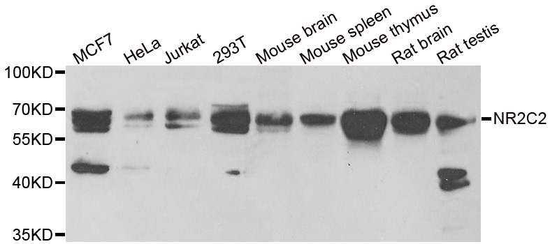
Figure 1. Western blot analysis of NR2C2 using anti-NR2C2 antibody (A02752-2). Electrophoresis was performed on a 5-20% SDS-PAGE gel at 70V (Stacking gel) / 90V (Resolving gel) for 2-3 hours. The sample well of each lane was loaded with 30 ug of sample under reducing conditions. Lane 1: human Hela whole cell lysates, Lane 2: human Jurkat whole cell lysates, Lane 3: human PC-3 whole cell lysates, Lane 4: human 293T whole cell lysates, Lane 5: monkey COS-7 whole cell lysates, Lane 6: human HEL whole cell lysates, Lane 7: human U20S whole cell lysates, Lane 8: human SH-SY5Y whole cell lysates, Lane 9: rat C6 whole cell lysates, Lane 10: mouse thymus tissue lysates, Lane 11: mouse NIH/3T3 whole cell lysates. After electrophoresis, proteins were transferred to a nitrocellulose membrane at 150 mA for 50-90 minutes. Blocked the membrane with 5% non-fat milk/TBS for 1.5 hour at RT. The membrane was incubated with rabbit anti-NR2C2 antigen affinity purified polyclonal antibody (Catalog # A02752-2) at 0.5 microg/mL overnight at 4°C, then washed with TBS-0.1%Tween 3 times with 5 minutes each and probed with a goat anti-rabbit IgG-HRP secondary antibody at a dilution of 1:5000 for 1.5 hour at RT. The signal is developed using an Enhanced Chemiluminescent detection (ECL) kit (Catalog # EK1002) with Tanon 5200 system. A specific band was detected for NR2C2 at approximately 68 kDa. The expected band size for NR2C2 is at 68 kDa.
. NR2C2 was detected in a paraffin-embedded section of human acinic cell carcinoma parotid tissue. Heat mediated antigen retrieval was performed in EDTA buffer (pH 8.0, epitope retrieval solution). The tissue section was blocked with 10% goat serum. The tissue section was then incubated with 2 microg/ml rabbit anti-NR2C2 Antibody (A02752-2) overnight at 4°C. Peroxidase Conjugated Goat Anti-rabbit IgG was used as secondary antibody and incubated for 30 minutes at 37°C. The tissue section was developed using HRP Conjugated Rabbit IgG Super Vision Assay Kit (Catalog # SV0002) with DAB as the chromogen.")
. NR2C2 was detected in a paraffin-embedded section of human colorectal adenocarcinoma tissue. Heat mediated antigen retrieval was performed in EDTA buffer (pH 8.0, epitope retrieval solution). The tissue section was blocked with 10% goat serum. The tissue section was then incubated with 2 microg/ml rabbit anti-NR2C2 Antibody (A02752-2) overnight at 4°C. Peroxidase Conjugated Goat Anti-rabbit IgG was used as secondary antibody and incubated for 30 minutes at 37°C. The tissue section was developed using HRP Conjugated Rabbit IgG Super Vision Assay Kit (Catalog # SV0002) with DAB as the chromogen.")
. Overlay histogram showing 293T cells stained with A02752-2 (Blue line). To facilitate intracellular staining, cells were fixed with 4% paraformaldehyde and permeabilized with permeabilization buffer. The cells were blocked with 10% normal goat serum. And then incubated with rabbit anti-NR2C2 Antibody (A02752-2, 1 microg/1x106 cells) for 30 min at 20°C. DyLight®488 conjugated goat anti-rabbit IgG (BA1127, 5-10 microg/1x106 cells) was used as secondary antibody for 30 minutes at 20°C. Isotype control antibody (Green line) was rabbit IgG (1 microg/1x106) used under the same conditions. Unlabelled sample (Red line) was also used as a control.")
Figure 1. Western blot analysis of NR2C2 using anti-NR2C2 antibody (A02752-2). Electrophoresis was performed on a 5-20% SDS-PAGE gel at 70V (Stacking gel) / 90V (Resolving gel) for 2-3 hours. The sample well of each lane was loaded with 30 ug of sample under reducing conditions. Lane 1: human Hela whole cell lysates, Lane 2: human Jurkat whole cell lysates, Lane 3: human PC-3 whole cell lysates, Lane 4: human 293T whole cell lysates, Lane 5: monkey COS-7 whole cell lysates, Lane 6: human HEL whole cell lysates, Lane 7: human U20S whole cell lysates, Lane 8: human SH-SY5Y whole cell lysates, Lane 9: rat C6 whole cell lysates, Lane 10: mouse thymus tissue lysates, Lane 11: mouse NIH/3T3 whole cell lysates. After electrophoresis, proteins were transferred to a nitrocellulose membrane at 150 mA for 50-90 minutes. Blocked the membrane with 5% non-fat milk/TBS for 1.5 hour at RT. The membrane was incubated with rabbit anti-NR2C2 antigen affinity purified polyclonal antibody (Catalog # A02752-2) at 0.5 microg/mL overnight at 4°C, then washed with TBS-0.1%Tween 3 times with 5 minutes each and probed with a goat anti-rabbit IgG-HRP secondary antibody at a dilution of 1:5000 for 1.5 hour at RT. The signal is developed using an Enhanced Chemiluminescent detection (ECL) kit (Catalog # EK1002) with Tanon 5200 system. A specific band was detected for NR2C2 at approximately 68 kDa. The expected band size for NR2C2 is at 68 kDa.
Anti-NR2C2 Antibody Picoband(r)
A02752-2-CARRIER-FREE
ApplicationsFlow Cytometry, Western Blot, ELISA, ImmunoHistoChemistry
Product group Antibodies
ReactivityHuman, Monkey, Mouse, Rat
TargetNR2C2
Overview
- SupplierBoster Bio
- Product NameAnti-NR2C2 Antibody Picoband(r)
- Delivery Days Customer9
- ApplicationsFlow Cytometry, Western Blot, ELISA, ImmunoHistoChemistry
- CertificationResearch Use Only
- ClonalityPolyclonal
- Concentration500 ug/ml
- Gene ID7182
- Target nameNR2C2
- Target descriptionnuclear receptor subfamily 2 group C member 2
- Target synonymsTAK1, TR4, nuclear receptor subfamily 2 group C member 2, Nuclear hormone receptor TR4, orphan nuclear receptor TAK1, orphan nuclear receptor TR4, orphan receptor TR4, testicular nuclear receptor 4
- HostRabbit
- IsotypeIgG
- Protein IDP49116
- Protein NameNuclear receptor subfamily 2 group C member 2
- Scientific DescriptionBoster Bio Anti-NR2C2 Antibody Picoband® catalog # A02752-2. Tested in ELISA, IHC, WB, Flow Cytometry applications. This antibody reacts with Human, Mouse, Rat, Monkey. The brand Picoband indicates this is a premium antibody that guarantees superior quality, high affinity, and strong signals with minimal background in Western blot applications. Only our best-performing antibodies are designated as Picoband, ensuring unmatched performance.
- ReactivityHuman, Monkey, Mouse, Rat
- Storage Instruction-20°C,2°C to 8°C
- UNSPSC12352203
Related products
Product group Antibodies
Anti-NR2C2 AntibodyA43386
ApplicationsWestern Blot
ReactivityHuman, Rat
- SizePrice
Product group Antibodies
Anti-NR2C2 Antibody144-66141
ApplicationsWestern Blot
ReactivityHuman, Mouse, Rat
TargetNR2C2
- SizePrice

Product group Antibodies
NR2C2 AntibodyCSB-PA016052ESR1HU
ApplicationsELISA, ImmunoHistoChemistry
ReactivityHuman
TargetNR2C2
- SizePrice
Product group Antibodies
ApplicationsImmunoPrecipitation, Western Blot, ImmunoCytoChemistry, ImmunoHistoChemistry
ReactivityMouse, Rat
TargetNR2C2
- SizePrice
Product group Antibodies
NR2C2 antibodyGTX105119
ApplicationsImmunoFluorescence, Western Blot, ImmunoCytoChemistry, ImmunoHistoChemistry, ImmunoHistoChemistry Paraffin
ReactivityHuman
TargetNR2C2
- SizePrice

Product group Antibodies
Anti-NR2C2 AntibodyHPA006313
ApplicationsWestern Blot, ChIP Chromatin ImmunoPrecipitation, ImmunoCytoChemistry, ImmunoHistoChemistry
ReactivityHuman, Mouse, Rat
TargetNR2C2
- SizePrice
Product group Antibodies

NR2C2 / TAK1 AntibodyLS-C334707
ApplicationsWestern Blot, ImmunoHistoChemistry
ReactivityHuman, Mouse, Rat
TargetNR2C2
- SizePrice
Product group Antibodies
References
ApplicationsFlow Cytometry, ImmunoFluorescence, Western Blot, ELISA, ImmunoCytoChemistry, ImmunoHistoChemistry, ImmunoHistoChemistry Frozen, ImmunoHistoChemistry Paraffin
ReactivityBovine, Chicken, Equine, Human, Mouse, Porcine, Rabbit, Rat
TargetNR2C2
- SizePrice
Product group Antibodies
Anti-NR2C2 AntibodyCAB6422
ApplicationsWestern Blot, ELISA
ReactivityHuman
- SizePrice