


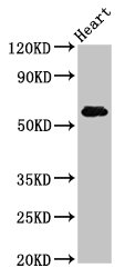
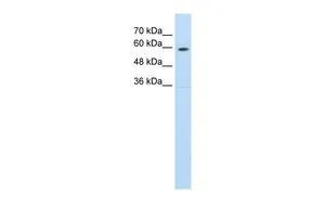
Figure 1. Western blot analysis of SLC11A1 using anti-SLC11A1 antibody (A02547-3). Electrophoresis was performed on a 5-20% SDS-PAGE gel at 70V (Stacking gel) / 90V (Resolving gel) for 2-3 hours. The sample well of each lane was loaded with 50ug of sample under reducing conditions. Lane 1: human K562 whole cell lysates Lane 2: human THP-1 whole cell lysates Lane 3: human placenta tissue lysates Lane 4: human A549 whole cell lysates Lane 5: human Caco-2 whole cell lysates Lane 6: human HepG2 whole cell lysates Lane 7: human U2OS whole cell lysates Lane 8: human Hela whole cell lysates After Electrophoresis, proteins were transferred to a Nitrocellulose membrane at 150mA for 50-90 minutes. Blocked the membrane with 5% Non-fat Milk/ TBS for 1.5 hour at RT. The membrane was incubated with rabbit anti-SLC11A1 antigen affinity purified polyclonal antibody (Catalog # A02547-3) at 0.5 microg/mL overnight at 4°C, then washed with TBS-0.1%Tween 3 times with 5 minutes each and probed with a goat anti-rabbit IgG-HRP secondary antibody at a dilution of 1:10000 for 1.5 hour at RT. The signal is developed using an Enhanced Chemiluminescent detection (ECL) kit (Catalog # EK1002) with Tanon 5200 system. A specific band was detected for SLC11A1 at approximately 90-120KD. The expected band size for SLC11A1 is at 60KD.
. SLC11A1 was detected in paraffin-embedded section of human intestinal cancer tissues. Heat mediated antigen retrieval was performed in citrate buffer (pH6, epitope retrieval solution) for 20 mins. The tissue section was blocked with 10% goat serum. The tissue section was then incubated with 1microg/ml rabbit anti-SLC11A1 Antibody (A02547-3) overnight at 4°C. Biotinylated goat anti-rabbit IgG was used as secondary antibody and incubated for 30 minutes at 37°C. The tissue section was developed using Strepavidin-Biotin-Complex (SABC)(Catalog # SA1022) with DAB as the chromogen.")
. SLC11A1 was detected in paraffin-embedded section of mouse spleen tissues. Heat mediated antigen retrieval was performed in citrate buffer (pH6, epitope retrieval solution) for 20 mins. The tissue section was blocked with 10% goat serum. The tissue section was then incubated with 1microg/ml rabbit anti-SLC11A1 Antibody (A02547-3) overnight at 4°C. Biotinylated goat anti-rabbit IgG was used as secondary antibody and incubated for 30 minutes at 37°C. The tissue section was developed using Strepavidin-Biotin-Complex (SABC)(Catalog # SA1022) with DAB as the chromogen.")
. SLC11A1 was detected in paraffin-embedded section of rat spleen tissues. Heat mediated antigen retrieval was performed in citrate buffer (pH6, epitope retrieval solution) for 20 mins. The tissue section was blocked with 10% goat serum. The tissue section was then incubated with 1microg/ml rabbit anti-SLC11A1 Antibody (A02547-3) overnight at 4°C. Biotinylated goat anti-rabbit IgG was used as secondary antibody and incubated for 30 minutes at 37°C. The tissue section was developed using Strepavidin-Biotin-Complex (SABC)(Catalog # SA1022) with DAB as the chromogen.")
. SLC11A1 was detected in immunocytochemical section of MCF-7 cell. Enzyme antigen retrieval was performed using IHC enzyme antigen retrieval reagent (AR0022) for 15 mins. The cells were blocked with 10% goat serum. And then incubated with 2microg/mL rabbit anti-SLC11A1 Antibody (A02547-3) overnight at 4°C. DyLight®594 Conjugated Goat Anti-Rabbit IgG (BA1142) was used as secondary antibody at 1:100 dilution and incubated for 30 minutes at 37°C. The section was counterstained with DAPI. Visualize using a fluorescence microscope and filter sets appropriate for the label used.")
Figure 1. Western blot analysis of SLC11A1 using anti-SLC11A1 antibody (A02547-3). Electrophoresis was performed on a 5-20% SDS-PAGE gel at 70V (Stacking gel) / 90V (Resolving gel) for 2-3 hours. The sample well of each lane was loaded with 50ug of sample under reducing conditions. Lane 1: human K562 whole cell lysates Lane 2: human THP-1 whole cell lysates Lane 3: human placenta tissue lysates Lane 4: human A549 whole cell lysates Lane 5: human Caco-2 whole cell lysates Lane 6: human HepG2 whole cell lysates Lane 7: human U2OS whole cell lysates Lane 8: human Hela whole cell lysates After Electrophoresis, proteins were transferred to a Nitrocellulose membrane at 150mA for 50-90 minutes. Blocked the membrane with 5% Non-fat Milk/ TBS for 1.5 hour at RT. The membrane was incubated with rabbit anti-SLC11A1 antigen affinity purified polyclonal antibody (Catalog # A02547-3) at 0.5 microg/mL overnight at 4°C, then washed with TBS-0.1%Tween 3 times with 5 minutes each and probed with a goat anti-rabbit IgG-HRP secondary antibody at a dilution of 1:10000 for 1.5 hour at RT. The signal is developed using an Enhanced Chemiluminescent detection (ECL) kit (Catalog # EK1002) with Tanon 5200 system. A specific band was detected for SLC11A1 at approximately 90-120KD. The expected band size for SLC11A1 is at 60KD.
Anti-NRAMP1/SLC11A1 Antibody Picoband(r)
A02547-3-CARRIER-FREE
ApplicationsImmunoFluorescence, Western Blot, ELISA, ImmunoCytoChemistry, ImmunoHistoChemistry
Product group Antibodies
ReactivityHuman, Mouse, Rat
TargetSLC11A1
Overview
- SupplierBoster Bio
- Product NameAnti-NRAMP1/SLC11A1 Antibody Picoband(r)
- Delivery Days Customer9
- ApplicationsImmunoFluorescence, Western Blot, ELISA, ImmunoCytoChemistry, ImmunoHistoChemistry
- CertificationResearch Use Only
- ClonalityPolyclonal
- Concentration500 ug/ml
- Gene ID6556
- Target nameSLC11A1
- Target descriptionsolute carrier family 11 member 1
- Target synonymsLSH, NRAMP, NRAMP1, natural resistance-associated macrophage protein 1, Leishmaniasis resistance, NRAMP 1, solute carrier family 11 (proton-coupled divalent metal ion transporter), member 1, solute carrier family 11 (proton-coupled divalent metal ion transporters), member 1, solute carrier family 11 (sodium/phosphate symporters), member 1
- HostRabbit
- IsotypeIgG
- Protein IDP49279
- Protein NameNatural resistance-associated macrophage protein 1
- Scientific DescriptionBoster Bio Anti-NRAMP1/SLC11A1 Antibody Picoband® catalog # A02547-3. Tested in ELISA, IF, IHC, ICC, WB applications. This antibody reacts with Human, Mouse, Rat. The brand Picoband indicates this is a premium antibody that guarantees superior quality, high affinity, and strong signals with minimal background in Western blot applications. Only our best-performing antibodies are designated as Picoband, ensuring unmatched performance.
- ReactivityHuman, Mouse, Rat
- Storage Instruction-20°C,2°C to 8°C
- UNSPSC12352203
Related products
Product group Antibodies

SLC11A1 / NRAMP AntibodyLS-C670766
ApplicationsWestern Blot, ELISA, ImmunoHistoChemistry, ImmunoHistoChemistry Paraffin
ReactivityHuman
TargetSLC11A1
- SizePrice
Product group Antibodies
SLC11A1 Polyclonal AntibodyCAC12925
ApplicationsImmunoFluorescence, Western Blot, ELISA, ImmunoHistoChemistry
ReactivityRat
TargetSLC11A1
- SizePrice
Product group Antibodies
SLC11A1 AntibodyCSB-PA021380LA01HU
ApplicationsImmunoFluorescence, Western Blot, ELISA, ImmunoHistoChemistry
ReactivityHuman, Rat
TargetSLC11A1
- SizePrice

Product group Antibodies
Anti-SLC11A1 AntibodyHPA029590
ApplicationsImmunoHistoChemistry
ReactivityHuman
TargetSLC11A1
- SizePrice
Product group Antibodies
SLC11A1 antibody, C-termGTX46862
ApplicationsWestern Blot
ReactivityHuman
TargetSLC11A1
- SizePrice
Product group Antibodies
SLC11A1 AntibodyPACO55778
ApplicationsImmunoFluorescence, Western Blot, ELISA, ImmunoHistoChemistry
ReactivityHuman, Rat
TargetSLC11A1
- SizePrice