


Anti-NUDT15 Antibody
A10407
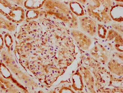
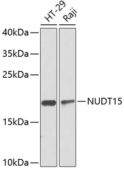
ApplicationsWestern Blot, ImmunoHistoChemistry
Product group Antibodies
ReactivityHuman, Mouse, Rat
Overview
- SupplierAntibodies.com
- Product NameAnti-NUDT15 Antibody
- Delivery Days Customer7
- ApplicationsWestern Blot, ImmunoHistoChemistry
- CertificationResearch Use Only
- ClonalityPolyclonal
- ConjugateUnconjugated
- HostRabbit
- IsotypeIgG
- Scientific DescriptionRabbit polyclonal antibody to NUDT15.
- ReactivityHuman, Mouse, Rat
- UNSPSC12352203
Related products
Product group Antibodies
NUDT15 AntibodyCSB-PA885733LA01HU
ApplicationsImmunoFluorescence, ELISA, ImmunoHistoChemistry
ReactivityHuman
TargetNUDT15
- SizePrice
Product group Antibodies
Anti-NUDT15 Antibody144-08368
ApplicationsWestern Blot, ImmunoHistoChemistry
ReactivityHuman
TargetNUDT15
- SizePrice
Product group Antibodies
NUDT15 antibodyGTX32759
ApplicationsWestern Blot, ImmunoHistoChemistry, ImmunoHistoChemistry Paraffin
ReactivityHuman
TargetNUDT15
- SizePrice

Product group Antibodies
Anti-NUDT15 Antibody Picoband(r)A05498-2-CARRIER-FREE
ApplicationsImmunoFluorescence, Western Blot, ELISA, ImmunoCytoChemistry
ReactivityHuman
TargetNUDT15
- SizePrice
Product group Antibodies

NUDT15 AntibodyLS-C749152
ApplicationsWestern Blot
ReactivityHuman, Mouse, Rat
TargetNUDT15
- SizePrice

Product group Antibodies
Anti-NUDT15 AntibodyHPA038969
ApplicationsImmunoHistoChemistry
ReactivityHuman
TargetNUDT15
- SizePrice