


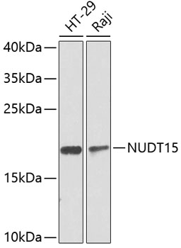
Figure 1. Western blot analysis of NUDT15 using anti-NUDT15 antibody (A05498-2). Electrophoresis was performed on a 5-20% SDS-PAGE gel at 70V (Stacking gel) / 90V (Resolving gel) for 2-3 hours. The sample well of each lane was loaded with 30 ug of sample under reducing conditions. Lane 1: human Jurkat whole cell lysates, Lane 2: human Hela whole cell lysates, Lane 3: human Caco-2 whole cell lysates. After electrophoresis, proteins were transferred to a nitrocellulose membrane at 150 mA for 50-90 minutes. Blocked the membrane with 5% non-fat milk/TBS for 1.5 hour at RT. The membrane was incubated with rabbit anti-NUDT15 antigen affinity purified polyclonal antibody (Catalog # A05498-2) at 0.5 microg/mL overnight at 4°C, then washed with TBS-0.1%Tween 3 times with 5 minutes each and probed with a goat anti-rabbit IgG-HRP secondary antibody at a dilution of 1:5000 for 1.5 hour at RT. The signal is developed using an Enhanced Chemiluminescent detection (ECL) kit (Catalog # EK1002) with Tanon 5200 system. A specific band was detected for NUDT15 at approximately 19 kDa. The expected band size for NUDT15 is at 19 kDa.
. NUDT15 was detected in an immunocytochemical section of A549 cells. Enzyme antigen retrieval was performed using IHC enzyme antigen retrieval reagent (AR0022) for 15 mins. The cells were blocked with 10% goat serum. And then incubated with 5 microg/mL rabbit anti-NUDT15 Antibody (A05498-2) overnight at 4°C. Cy3 Conjugated Goat Anti-Rabbit IgG (BA1032) was used as secondary antibody at 1:100 dilution and incubated for 30 minutes at 37°C. The tissue section was developed using Phalloidin-iFluor 488 Conjugated. Visualize using a fluorescence microscope and filter sets appropriate for the label used.")
Figure 1. Western blot analysis of NUDT15 using anti-NUDT15 antibody (A05498-2). Electrophoresis was performed on a 5-20% SDS-PAGE gel at 70V (Stacking gel) / 90V (Resolving gel) for 2-3 hours. The sample well of each lane was loaded with 30 ug of sample under reducing conditions. Lane 1: human Jurkat whole cell lysates, Lane 2: human Hela whole cell lysates, Lane 3: human Caco-2 whole cell lysates. After electrophoresis, proteins were transferred to a nitrocellulose membrane at 150 mA for 50-90 minutes. Blocked the membrane with 5% non-fat milk/TBS for 1.5 hour at RT. The membrane was incubated with rabbit anti-NUDT15 antigen affinity purified polyclonal antibody (Catalog # A05498-2) at 0.5 microg/mL overnight at 4°C, then washed with TBS-0.1%Tween 3 times with 5 minutes each and probed with a goat anti-rabbit IgG-HRP secondary antibody at a dilution of 1:5000 for 1.5 hour at RT. The signal is developed using an Enhanced Chemiluminescent detection (ECL) kit (Catalog # EK1002) with Tanon 5200 system. A specific band was detected for NUDT15 at approximately 19 kDa. The expected band size for NUDT15 is at 19 kDa.
Anti-NUDT15 Antibody Picoband(r)
A05498-2-CARRIER-FREE
ApplicationsImmunoFluorescence, Western Blot, ELISA, ImmunoCytoChemistry
Product group Antibodies
ReactivityHuman
TargetNUDT15
Overview
- SupplierBoster Bio
- Product NameAnti-NUDT15 Antibody Picoband(r)
- Delivery Days Customer9
- ApplicationsImmunoFluorescence, Western Blot, ELISA, ImmunoCytoChemistry
- CertificationResearch Use Only
- ClonalityPolyclonal
- Concentration500 ug/ml
- Gene ID55270
- Target nameNUDT15
- Target descriptionnudix hydrolase 15
- Target synonymsMTH2, NUDT15D, nucleotide triphosphate diphosphatase NUDT15, 8-oxo-dGTPase NUDT15, mutT homolog 2, nucleoside diphosphate-linked to another moiety X hydrolase 15, nudix (nucleoside diphosphate linked moiety X)-type motif 15, probable 7,8-dihydro-8-oxoguanine triphosphatase NUDT15, probable 8-oxo-dGTP diphosphatase NUDT15
- HostRabbit
- IsotypeIgG
- Protein IDQ9NV35
- Protein NameNucleotide triphosphate diphosphatase NUDT15
- Scientific DescriptionBoster Bio Anti-NUDT15 Antibody Picoband® catalog # A05498-2. Tested in ELISA, IF, ICC, WB applications. This antibody reacts with Human. The brand Picoband indicates this is a premium antibody that guarantees superior quality, high affinity, and strong signals with minimal background in Western blot applications. Only our best-performing antibodies are designated as Picoband, ensuring unmatched performance.
- ReactivityHuman
- Storage Instruction-20°C,2°C to 8°C
- UNSPSC12352203
Related products
Product group Antibodies
Anti-NUDT15 Antibody144-08368
ApplicationsWestern Blot, ImmunoHistoChemistry
ReactivityHuman
TargetNUDT15
- SizePrice
Product group Antibodies

NUDT15 AntibodyLS-C749152
ApplicationsWestern Blot
ReactivityHuman, Mouse, Rat
TargetNUDT15
- SizePrice
Product group Antibodies
Anti-NUDT15 AntibodyA10407
ApplicationsWestern Blot, ImmunoHistoChemistry
ReactivityHuman, Mouse, Rat
- SizePrice
Product group Antibodies
NUDT15 antibodyGTX32759
ApplicationsWestern Blot, ImmunoHistoChemistry, ImmunoHistoChemistry Paraffin
ReactivityHuman
TargetNUDT15
- SizePrice

Product group Antibodies
Anti-NUDT15 AntibodyHPA038969
ApplicationsImmunoHistoChemistry
ReactivityHuman
TargetNUDT15
- SizePrice
Product group Antibodies
NUDT15 AntibodyCSB-PA885733LA01HU
ApplicationsImmunoFluorescence, ELISA, ImmunoHistoChemistry
ReactivityHuman
TargetNUDT15
- SizePrice