


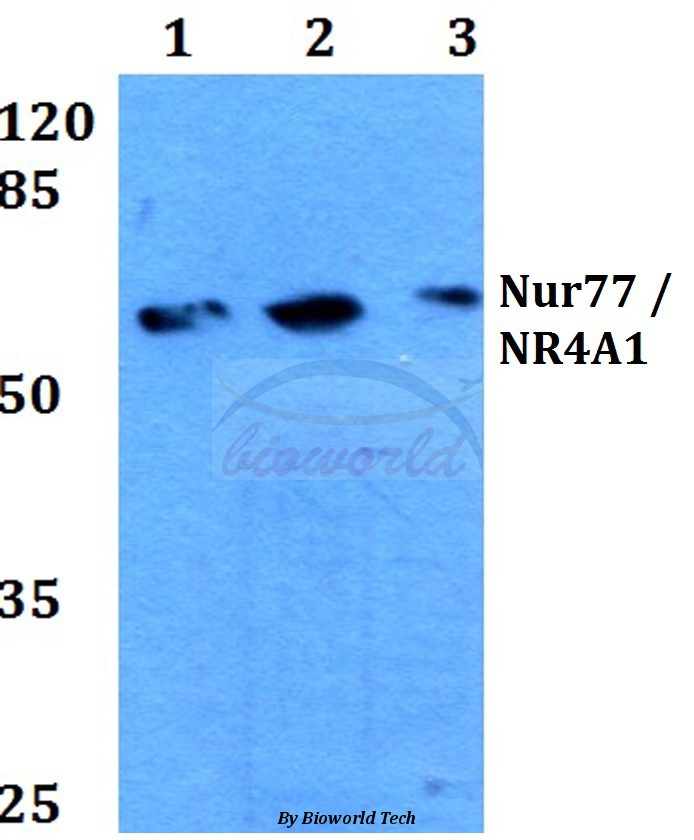
Figure 1. Western blot analysis of NUR77 using anti-NUR77 antibody (PB9766). Electrophoresis was performed on a 5-20% SDS-PAGE gel at 70V (Stacking gel) / 90V (Resolving gel) for 2-3 hours. Lane 1: Rat Brain Tissue Lysate at 50ug, Lane 2: Mouse Brain Tissue Lysate at 50ug, Lane 3: U87 Whole Cell Lysate at 40ug, Lane 4: HELA Whole Cell Lysate at 40ug. After electrophoresis, proteins were transferred to a nitrocellulose membrane at 150 mA for 50-90 minutes. Blocked the membrane with 5% non-fat milk/TBS for 1.5 hour at RT. The membrane was incubated with rabbit anti-NUR77 antigen affinity purified polyclonal antibody (Catalog # PB9766) at 0.5 microg/mL overnight at 4°C, then washed with TBS-0.1%Tween 3 times with 5 minutes each and probed with a goat anti-rabbit IgG-HRP secondary antibody at a dilution of 1:5000 for 1.5 hour at RT. The signal is developed using an Enhanced Chemiluminescent detection (ECL) kit (Catalog # EK1002) with Tanon 5200 system. A specific band was detected for NUR77 at approximately 67 kDa. The expected band size for NUR77 is at 67 kDa.
Figure 1. Western blot analysis of NUR77 using anti-NUR77 antibody (PB9766). Electrophoresis was performed on a 5-20% SDS-PAGE gel at 70V (Stacking gel) / 90V (Resolving gel) for 2-3 hours. Lane 1: Rat Brain Tissue Lysate at 50ug, Lane 2: Mouse Brain Tissue Lysate at 50ug, Lane 3: U87 Whole Cell Lysate at 40ug, Lane 4: HELA Whole Cell Lysate at 40ug. After electrophoresis, proteins were transferred to a nitrocellulose membrane at 150 mA for 50-90 minutes. Blocked the membrane with 5% non-fat milk/TBS for 1.5 hour at RT. The membrane was incubated with rabbit anti-NUR77 antigen affinity purified polyclonal antibody (Catalog # PB9766) at 0.5 microg/mL overnight at 4°C, then washed with TBS-0.1%Tween 3 times with 5 minutes each and probed with a goat anti-rabbit IgG-HRP secondary antibody at a dilution of 1:5000 for 1.5 hour at RT. The signal is developed using an Enhanced Chemiluminescent detection (ECL) kit (Catalog # EK1002) with Tanon 5200 system. A specific band was detected for NUR77 at approximately 67 kDa. The expected band size for NUR77 is at 67 kDa.
Anti-NUR77/NR4A1 Antibody Picoband(r)
PB9766-CARRIER-FREE
ApplicationsWestern Blot
Product group Antibodies
ReactivityHuman, Mouse, Rat
TargetNR4A1
Overview
- SupplierBoster Bio
- Product NameAnti-NUR77/NR4A1 Antibody Picoband(r)
- Delivery Days Customer9
- Application Supplier NoteTested Species: In-house tested species with positive results. Other applications have not been tested. Optimal dilutions should be determined by end users.
- ApplicationsWestern Blot
- CertificationResearch Use Only
- ClonalityPolyclonal
- Concentration500 ug/ml
- Gene ID3164
- Target nameNR4A1
- Target descriptionnuclear receptor subfamily 4 group A member 1
- Target synonymsGFRP1, HMR, N10, NAK-1, NGFIB, NP10, NUR77, TR3, nuclear receptor subfamily 4immunitygroup A member 1, ST-59, TR3 orphan receptor, early response protein NAK1, growth factor-inducible nuclear protein N10, hormone receptor, nuclear hormone receptor NUR/77, orphan nuclear receptor HMR, orphan nuclear receptor TR3, steroid receptor TR3, testicular receptor 3
- HostRabbit
- IsotypeIgG
- Protein IDP22736
- Protein NameNuclear receptor subfamily 4immunitygroup A member 1
- Scientific DescriptionBoster Bio Anti-NUR77/NR4A1 Antibody Picoband® catalog # PB9766. Tested in WB applications. This antibody reacts with Human, Mouse, Rat. The brand Picoband indicates this is a premium antibody that guarantees superior quality, high affinity, and strong signals with minimal background in Western blot applications. Only our best-performing antibodies are designated as Picoband, ensuring unmatched performance.
- ReactivityHuman, Mouse, Rat
- Storage Instruction-20°C,2°C to 8°C
- UNSPSC12352203
Related products
Product group Antibodies
NR4A1 AntibodyCSB-PA003493
ApplicationsWestern Blot, ELISA, ImmunoHistoChemistry
ReactivityHuman, Monkey, Mouse, Rat
TargetNR4A1
- SizePrice
Product group Antibodies

ApplicationsELISA, ImmunoHistoChemistry
- SizePrice
Product group Antibodies
ApplicationsWestern Blot, ImmunoHistoChemistry
ReactivityHuman, Mouse, Rat
- SizePrice
Product group Antibodies

NR4A1 / NUR77 AntibodyLS-C746761
ApplicationsImmunoFluorescence, Western Blot
ReactivityHuman
TargetNR4A1
- SizePrice
Product group Antibodies

ApplicationsWestern Blot, ELISA, ImmunoHistoChemistry
ReactivityCanine, Human, Mouse, Rat
TargetNR4A1
- SizePrice

Product group Antibodies
Anti-NR4A1 AntibodyHPA070142
ApplicationsImmunoHistoChemistry
ReactivityHuman
TargetNR4A1
- SizePrice
Product group Antibodies
Nur77 Polyclonal AntibodyBS-3513R
ApplicationsFlow Cytometry, ImmunoFluorescence, Western Blot, ELISA, ImmunoCytoChemistry, ImmunoHistoChemistry, ImmunoHistoChemistry Frozen, ImmunoHistoChemistry Paraffin
ReactivityHuman, Mouse, Rat
TargetNR4A1
- SizePrice
Product group Antibodies
NUR77 antibodyGTX100797
ApplicationsWestern Blot, ImmunoHistoChemistry, ImmunoHistoChemistry Paraffin
ReactivityHuman, Mouse
TargetNR4A1
- SizePrice
Product group Antibodies
Anti-NR4A1Y058287
ApplicationsWestern Blot, ELISA, ImmunoHistoChemistry
ReactivityHuman, Mouse, Rat
- SizePrice