


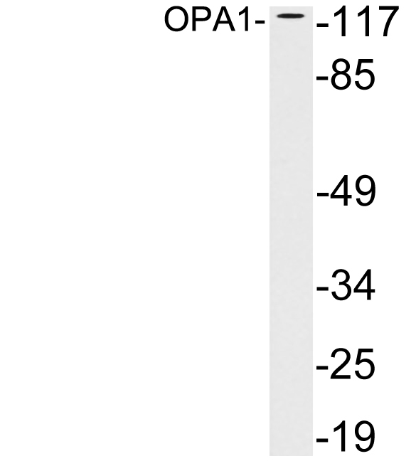
Figure 1. Western blot analysis of OPA1 using anti-OPA1 antibody (A00508-1). Electrophoresis was performed on a 5-20% SDS-PAGE gel at 70V (Stacking gel) / 90V (Resolving gel) for 2-3 hours. The sample well of each lane was loaded with 30 ug of sample under reducing conditions. Lane 1: human Ramos whole cell lysates, Lane 2: human Hela whole cell lysates, Lane 3: human PANC-1 whole cell lysates, Lane 4: human MCF-7 whole cell lysates, Lane 5: rat brain tissue lysates, Lane 6: mouse eye tissue lysates, Lane 7: mouse brain tissue lysates. After electrophoresis, proteins were transferred to a nitrocellulose membrane at 150 mA for 50-90 minutes. Blocked the membrane with 5% non-fat milk/TBS for 1.5 hour at RT. The membrane was incubated with rabbit anti-OPA1 antigen affinity purified polyclonal antibody (Catalog # A00508-1) at 0.25 microg/mL overnight at 4°C, then washed with TBS-0.1%Tween 3 times with 5 minutes each and probed with a goat anti-rabbit IgG-HRP secondary antibody at a dilution of 1:5000 for 1.5 hour at RT. The signal is developed using an Enhanced Chemiluminescent detection (ECL) kit (Catalog # EK1002) with Tanon 5200 system. A specific band was detected for OPA1 at approximately 80-100 kDa. The expected band size for OPA1 is at 112,80-100 kDa.
. OPA1 was detected in an immunocytochemical section of U2OS cells. Enzyme antigen retrieval was performed using IHC enzyme antigen retrieval reagent (AR0022) for 15 mins. The cells were blocked with 10% goat serum. And then incubated with 5 microg/mL rabbit anti-OPA1 Antibody (A00508-1) overnight at 4°C. Cy3 Conjugated Goat Anti-Rabbit IgG (BA1032) was used as secondary antibody at 1:500 dilution and incubated for 30 minutes at 37°C. The section was counterstained with DAPI. Visualize using a fluorescence microscope and filter sets appropriate for the label used.")
. Overlay histogram showing Daudi cells stained with A00508-1 (Blue line). To facilitate intracellular staining, cells were fixed with 4% paraformaldehyde and permeabilized with permeabilization buffer. The cells were blocked with 10% normal goat serum. And then incubated with rabbit anti-OPA1 Antibody (A00508-1, 1 microg/1x106 cells) for 30 min at 20°C. DyLight®488 conjugated goat anti-rabbit IgG (BA1127, 5-10 microg/1x106 cells) was used as secondary antibody for 30 minutes at 20°C. Isotype control antibody (Green line) was rabbit IgG (1 microg/1x106) used under the same conditions. Unlabelled sample without incubation with primary antibody and secondary antibody (Red line) was used as a blank control.")
Figure 1. Western blot analysis of OPA1 using anti-OPA1 antibody (A00508-1). Electrophoresis was performed on a 5-20% SDS-PAGE gel at 70V (Stacking gel) / 90V (Resolving gel) for 2-3 hours. The sample well of each lane was loaded with 30 ug of sample under reducing conditions. Lane 1: human Ramos whole cell lysates, Lane 2: human Hela whole cell lysates, Lane 3: human PANC-1 whole cell lysates, Lane 4: human MCF-7 whole cell lysates, Lane 5: rat brain tissue lysates, Lane 6: mouse eye tissue lysates, Lane 7: mouse brain tissue lysates. After electrophoresis, proteins were transferred to a nitrocellulose membrane at 150 mA for 50-90 minutes. Blocked the membrane with 5% non-fat milk/TBS for 1.5 hour at RT. The membrane was incubated with rabbit anti-OPA1 antigen affinity purified polyclonal antibody (Catalog # A00508-1) at 0.25 microg/mL overnight at 4°C, then washed with TBS-0.1%Tween 3 times with 5 minutes each and probed with a goat anti-rabbit IgG-HRP secondary antibody at a dilution of 1:5000 for 1.5 hour at RT. The signal is developed using an Enhanced Chemiluminescent detection (ECL) kit (Catalog # EK1002) with Tanon 5200 system. A specific band was detected for OPA1 at approximately 80-100 kDa. The expected band size for OPA1 is at 112,80-100 kDa.
Anti-OPA1 Antibody Picoband(r)
A00508-1-CARRIER-FREE
ApplicationsFlow Cytometry, ImmunoFluorescence, Western Blot, ELISA, ImmunoCytoChemistry
Product group Antibodies
ReactivityHuman, Mouse, Rat
TargetOPA1
Overview
- SupplierBoster Bio
- Product NameAnti-OPA1 Antibody Picoband(r)
- Delivery Days Customer9
- ApplicationsFlow Cytometry, ImmunoFluorescence, Western Blot, ELISA, ImmunoCytoChemistry
- CertificationResearch Use Only
- ClonalityPolyclonal
- Concentration500 ug/ml
- Gene ID4976
- Target nameOPA1
- Target descriptionOPA1 mitochondrial dynamin like GTPase
- Target synonymsBERHS, MGM1, MTDPS14, NPG, NTG, largeG, dynamin-like GTPase OPA1, mitochondrial, dynamin-like 120 kDa protein, mitochondrial, dynamin-like guanosine triphosphatase, mitochondrial dynamin-like GTPase, optic atrophy 1 (autosomal dominant), optic atrophy protein 1
- HostRabbit
- IsotypeIgG
- Protein IDO60313
- Protein NameDynamin-like GTPase OPA1, mitochondrial
- Scientific DescriptionBoster Bio Anti-OPA1 Antibody Picoband® catalog # A00508-1. Tested in ELISA, Flow Cytometry, IF, ICC, WB applications. This antibody reacts with Human, Mouse, Rat. The brand Picoband indicates this is a premium antibody that guarantees superior quality, high affinity, and strong signals with minimal background in Western blot applications. Only our best-performing antibodies are designated as Picoband, ensuring unmatched performance.
- ReactivityHuman, Mouse, Rat
- Storage Instruction-20°C,2°C to 8°C
- UNSPSC12352203
Related products
Product group Antibodies
Anti-OPA1 AntibodyA97989
ApplicationsWestern Blot, ELISA
ReactivityHuman, Mouse, Rat
- SizePrice
Product group Antibodies
Anti-OPA1 Antibody144-09833
ApplicationsImmunoFluorescence, Western Blot
ReactivityHuman, Mouse, Rat
TargetOPA1
- SizePrice
Product group Antibodies
OPA1 Polyclonal AntibodyCAC13144
ApplicationsWestern Blot, ELISA
ReactivityMouse
TargetOPA1
- SizePrice
Product group Antibodies
OPA1 AntibodyCSB-PA016340HA01HU
ApplicationsELISA, ImmunoHistoChemistry
ReactivityHuman
TargetOPA1
- SizePrice
Product group Antibodies

OPA1 AntibodyLS-C498037
ApplicationsWestern Blot
ReactivityHuman, Mouse, Rat
TargetOPA1
- SizePrice
Product group Antibodies
OPA1 antibodyGTX129917
ApplicationsWestern Blot
ReactivityHuman, Mouse, Rat
TargetOPA1
- SizePrice

Product group Antibodies
Anti-OPA1 AntibodyHPA036926
ApplicationsWestern Blot, ImmunoHistoChemistry
ReactivityHuman
TargetOPA1
- SizePrice
Product group Antibodies
Anti-OPA1 AntibodyCAB9833
ApplicationsImmunoFluorescence, Western Blot, ELISA, ImmunoCytoChemistry
ReactivityHuman
TargetOPA1
- SizePrice