

IHC image of CSB-PA016340HA01HU diluted at 1:300 and staining in paraffin-embedded human small intestine tissue performed on a Leica BondTM system. After dewaxing and hydration, antigen retrieval was mediated by high pressure in a citrate buffer (pH 6.0). Section was blocked with 10% normal goat serum 30min at RT. Then primary antibody (1% BSA) was incubated at 4°C overnight. The primary is detected by a biotinylated secondary antibody and visualized using an HRP conjugated SP system.
. Section was blocked with 10% normal goat serum 30min at RT. Then primary antibody (1% BSA) was incubated at 4°C overnight. The primary is detected by a biotinylated secondary antibody and visualized using an HRP conjugated SP system.")
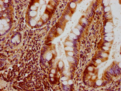
IHC image of CSB-PA016340HA01HU diluted at 1:300 and staining in paraffin-embedded human small intestine tissue performed on a Leica BondTM system. After dewaxing and hydration, antigen retrieval was mediated by high pressure in a citrate buffer (pH 6.0). Section was blocked with 10% normal goat serum 30min at RT. Then primary antibody (1% BSA) was incubated at 4°C overnight. The primary is detected by a biotinylated secondary antibody and visualized using an HRP conjugated SP system.
OPA1 Antibody
CSB-PA016340HA01HU
ApplicationsELISA, ImmunoHistoChemistry
Product group Antibodies
ReactivityHuman
TargetOPA1
Overview
- SupplierCusabio
- Product NameOPA1 Antibody
- Delivery Days Customer20
- ApplicationsELISA, ImmunoHistoChemistry
- CertificationResearch Use Only
- ClonalityPolyclonal
- ConjugateUnconjugated
- Gene ID4976
- Target nameOPA1
- Target descriptionOPA1 mitochondrial dynamin like GTPase
- Target synonymsBERHS, MGM1, MTDPS14, NPG, NTG, largeG, dynamin-like GTPase OPA1, mitochondrial, dynamin-like 120 kDa protein, mitochondrial, dynamin-like guanosine triphosphatase, mitochondrial dynamin-like GTPase, optic atrophy 1 (autosomal dominant), optic atrophy protein 1
- HostRabbit
- IsotypeIgG
- Protein IDO60313
- Protein NameDynamin-like GTPase OPA1, mitochondrial
- Scientific DescriptionDynamin-related GTPase required for mitochondrial fusion and regulation of apoptosis. May form a diffusion barrier for proteins stored in mitochondrial cristae. Proteolytic processing in response to intrinsic apoptotic signals may lead to disassembly of OPA1 oligomers and release of the caspase activator cytochrome C (CYCS) into the mitochondrial intermembrane space. May also play a role in mitochondrial genome maintenance.
- ReactivityHuman
- Storage Instruction-20°C or -80°C
- UNSPSC41116161
Related products
Product group Antibodies
Anti-OPA1 AntibodyA97989
ApplicationsWestern Blot, ELISA
ReactivityHuman, Mouse, Rat
- SizePrice
Product group Antibodies
Anti-OPA1 Antibody144-09833
ApplicationsImmunoFluorescence, Western Blot
ReactivityHuman, Mouse, Rat
TargetOPA1
- SizePrice

Product group Antibodies
Anti-OPA1 Antibody Picoband(r)A00508-1-CARRIER-FREE
ApplicationsFlow Cytometry, ImmunoFluorescence, Western Blot, ELISA, ImmunoCytoChemistry
ReactivityHuman, Mouse, Rat
TargetOPA1
- SizePrice
Product group Antibodies
OPA1 Polyclonal AntibodyCAC13144
ApplicationsWestern Blot, ELISA
ReactivityMouse
TargetOPA1
- SizePrice
Product group Antibodies

OPA1 AntibodyLS-C498037
ApplicationsWestern Blot
ReactivityHuman, Mouse, Rat
TargetOPA1
- SizePrice
Product group Antibodies
OPA1 antibodyGTX129917
ApplicationsWestern Blot
ReactivityHuman, Mouse, Rat
TargetOPA1
- SizePrice

Product group Antibodies
Anti-OPA1 AntibodyHPA036926
ApplicationsWestern Blot, ImmunoHistoChemistry
ReactivityHuman
TargetOPA1
- SizePrice
Product group Antibodies
Anti-OPA1 AntibodyCAB9833
ApplicationsImmunoFluorescence, Western Blot, ELISA, ImmunoCytoChemistry
ReactivityHuman
TargetOPA1
- SizePrice