


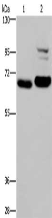
Figure 1. Western blot analysis of OS9 using anti-OS9 antibody (A04158-1). Electrophoresis was performed on a 5-20% SDS-PAGE gel at 70V (Stacking gel) / 90V (Resolving gel) for 2-3 hours. The sample well of each lane was loaded with 30 ug of sample under reducing conditions. Lane 1: human Hela whole cell lysates, Lane 2: human 293T whole cell lysates, Lane 3: human HL-60 whole cell lysates, Lane 4: rat pancreas tissue lysates, Lane 5: mouse pancreas tissue lysates. After electrophoresis, proteins were transferred to a nitrocellulose membrane at 150 mA for 50-90 minutes. Blocked the membrane with 5% non-fat milk/TBS for 1.5 hour at RT. The membrane was incubated with rabbit anti-OS9 antigen affinity purified polyclonal antibody (Catalog # A04158-1) at 0.5 microg/mL overnight at 4°C, then washed with TBS-0.1%Tween 3 times with 5 minutes each and probed with a goat anti-rabbit IgG-HRP secondary antibody at a dilution of 1:5000 for 1.5 hour at RT. The signal is developed using an Enhanced Chemiluminescent detection (ECL) kit (Catalog # EK1002) with Tanon 5200 system. A specific band was detected for OS9 at approximately 83 kDa. The expected band size for OS9 is at 76 kDa.
. OS9 was detected in an immunocytochemical section of Siha cells. Enzyme antigen retrieval was performed using IHC enzyme antigen retrieval reagent (AR0022) for 15 mins. The cells were blocked with 10% goat serum. And then incubated with 5 microg/mL rabbit anti-OS9 Antibody (A04158-1) overnight at 4°C. DyLight®488 Conjugated Goat Anti-Rabbit IgG (BA1127) was used as secondary antibody at 1:100 dilution and incubated for 30 minutes at 37°C. The section was counterstained with DAPI. Visualize using a fluorescence microscope and filter sets appropriate for the label used.")
Figure 1. Western blot analysis of OS9 using anti-OS9 antibody (A04158-1). Electrophoresis was performed on a 5-20% SDS-PAGE gel at 70V (Stacking gel) / 90V (Resolving gel) for 2-3 hours. The sample well of each lane was loaded with 30 ug of sample under reducing conditions. Lane 1: human Hela whole cell lysates, Lane 2: human 293T whole cell lysates, Lane 3: human HL-60 whole cell lysates, Lane 4: rat pancreas tissue lysates, Lane 5: mouse pancreas tissue lysates. After electrophoresis, proteins were transferred to a nitrocellulose membrane at 150 mA for 50-90 minutes. Blocked the membrane with 5% non-fat milk/TBS for 1.5 hour at RT. The membrane was incubated with rabbit anti-OS9 antigen affinity purified polyclonal antibody (Catalog # A04158-1) at 0.5 microg/mL overnight at 4°C, then washed with TBS-0.1%Tween 3 times with 5 minutes each and probed with a goat anti-rabbit IgG-HRP secondary antibody at a dilution of 1:5000 for 1.5 hour at RT. The signal is developed using an Enhanced Chemiluminescent detection (ECL) kit (Catalog # EK1002) with Tanon 5200 system. A specific band was detected for OS9 at approximately 83 kDa. The expected band size for OS9 is at 76 kDa.
Anti-OS9 Antibody Picoband(r)
A04158-1-CARRIER-FREE
ApplicationsImmunoFluorescence, Western Blot, ELISA, ImmunoCytoChemistry
Product group Antibodies
ReactivityHuman, Mouse, Rat
TargetOS9
Overview
- SupplierBoster Bio
- Product NameAnti-OS9 Antibody Picoband(r)
- Delivery Days Customer9
- ApplicationsImmunoFluorescence, Western Blot, ELISA, ImmunoCytoChemistry
- CertificationResearch Use Only
- ClonalityPolyclonal
- Concentration500 ug/ml
- Gene ID10956
- Target nameOS9
- Target descriptionOS9 endoplasmic reticulum lectin
- Target synonymsERLEC2, OS-9, protein OS-9, amplified in osteosarcoma 9, endoplasmic reticulum lectin 2, erlectin 2, osteosarcoma amplified 9, endoplasmic reticulum associated protein, osteosarcoma amplified 9, endoplasmic reticulum lectin
- HostRabbit
- IsotypeIgG
- Protein IDQ13438
- Protein NameProtein OS-9
- Scientific DescriptionBoster Bio Anti-OS9 Antibody Picoband® catalog # A04158-1. Tested in ELISA, IF, ICC, WB applications. This antibody reacts with Human, Mouse, Rat. The brand Picoband indicates this is a premium antibody that guarantees superior quality, high affinity, and strong signals with minimal background in Western blot applications. Only our best-performing antibodies are designated as Picoband, ensuring unmatched performance.
- ReactivityHuman, Mouse, Rat
- Storage Instruction-20°C,2°C to 8°C
- UNSPSC12352203
Related products
Product group Antibodies
Anti-OS9 AntibodyA81203
ApplicationsWestern Blot
ReactivityHuman, Mouse
- SizePrice
Product group Antibodies
Anti-OS9 (C-term) Antibody102-22653
ApplicationsWestern Blot
TargetOS9
- SizePrice
Product group Antibodies
OS9 Recombinant Antibody, AbBy Fluor-405 ConjugatedBSM-61925R-BF405
ApplicationsImmunoFluorescence, Western Blot
ReactivityHuman, Mouse, Rat
TargetOS9
- SizePrice
Product group Antibodies
OS9 AntibodyCSB-PA577148
ApplicationsWestern Blot, ELISA
ReactivityHuman, Mouse
TargetOS9
- SizePrice
Product group Antibodies
OS9 Polyclonal AntibodyCAC14774
ApplicationsImmunoFluorescence, Western Blot, ELISA, ImmunoHistoChemistry
TargetOS9
- SizePrice
Product group Antibodies

OS9 AntibodyLS-C405219
ApplicationsWestern Blot, ELISA, ImmunoHistoChemistry
ReactivityHuman, Mouse
TargetOS9
- SizePrice

Product group Antibodies
Anti-OS9 AntibodyHPA013694
ApplicationsImmunoHistoChemistry
ReactivityHuman
TargetOS9
- SizePrice