


Immunohistochemical staining of human placenta shows cytoplasmic positivity in trophoblastic cells.
Immunohistochemical staining of human placenta shows cytoplasmic positivity in trophoblastic cells.
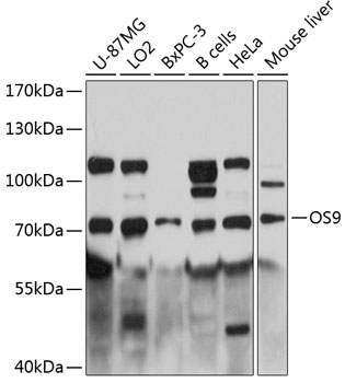
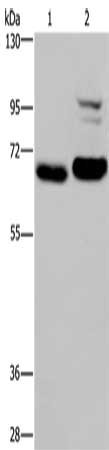
Anti-OS9 Antibody
HPA013694
ApplicationsImmunoHistoChemistry
Product group Antibodies
ReactivityHuman
TargetOS9
Overview
- SupplierAtlas Antibodies
- Product NameAnti-OS9 Antibody
- Delivery Days Customer4
- ApplicationsImmunoHistoChemistry
- CertificationResearch Use Only
- ClonalityPolyclonal
- ConjugateUnconjugated
- Gene ID10956
- Target nameOS9
- Target descriptionOS9 endoplasmic reticulum lectin
- Target synonymsERLEC2, OS-9, protein OS-9, amplified in osteosarcoma 9, endoplasmic reticulum lectin 2, erlectin 2, osteosarcoma amplified 9, endoplasmic reticulum associated protein, osteosarcoma amplified 9, endoplasmic reticulum lectin
- HostRabbit
- IsotypeIgG
- Protein IDQ13438
- Protein NameProtein OS-9
- Scientific DescriptionRecombinant Protein Epitope Signature Tag (PrEST) antigen sequence
- ReactivityHuman
- Storage Instruction-20°C,2°C to 8°C
- UNSPSC41116161
Datasheet
MSDS
Related products
Product group Antibodies
Anti-OS9 AntibodyA81203
ApplicationsWestern Blot
ReactivityHuman, Mouse
- SizePrice
Product group Antibodies
Anti-OS9 (C-term) Antibody102-22653
ApplicationsWestern Blot
TargetOS9
- SizePrice

Product group Antibodies
Anti-OS9 Antibody Picoband(r)A04158-1-CARRIER-FREE
ApplicationsImmunoFluorescence, Western Blot, ELISA, ImmunoCytoChemistry
ReactivityHuman, Mouse, Rat
TargetOS9
- SizePrice
Product group Antibodies
OS9 Recombinant Antibody, AbBy Fluor-405 ConjugatedBSM-61925R-BF405
ApplicationsImmunoFluorescence, Western Blot
ReactivityHuman, Mouse, Rat
TargetOS9
- SizePrice
Product group Antibodies
OS9 AntibodyCSB-PA577148
ApplicationsWestern Blot, ELISA
ReactivityHuman, Mouse
TargetOS9
- SizePrice
Product group Antibodies
OS9 Polyclonal AntibodyCAC14774
ApplicationsImmunoFluorescence, Western Blot, ELISA, ImmunoHistoChemistry
TargetOS9
- SizePrice
Product group Antibodies

OS9 AntibodyLS-C405219
ApplicationsWestern Blot, ELISA, ImmunoHistoChemistry
ReactivityHuman, Mouse
TargetOS9
- SizePrice