


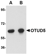
Figure 1. Western blot analysis of OTUD5 using anti-OTUD5 antibody (A09744-1). Electrophoresis was performed on a 5-20% SDS-PAGE gel at 70V (Stacking gel) / 90V (Resolving gel) for 2-3 hours. The sample well of each lane was loaded with 30 ug of sample under reducing conditions. Lane 1: human 293T whole cell lysates, Lane 2: human Hela whole cell lysates, Lane 3: human Jurkat whole cell lysates, Lane 4: human K562 whole cell lysates, Lane 5: rat liver tissue lysates, Lane 6: rat C6 whole cell lysates. After electrophoresis, proteins were transferred to a nitrocellulose membrane at 150 mA for 50-90 minutes. Blocked the membrane with 5% non-fat milk/TBS for 1.5 hour at RT. The membrane was incubated with rabbit anti-OTUD5 antigen affinity purified polyclonal antibody (Catalog # A09744-1) at 0.5 microg/mL overnight at 4°C, then washed with TBS-0.1%Tween 3 times with 5 minutes each and probed with a goat anti-rabbit IgG-HRP secondary antibody at a dilution of 1:5000 for 1.5 hour at RT. The signal is developed using an Enhanced Chemiluminescent detection (ECL) kit (Catalog # EK1002) with Tanon 5200 system. A specific band was detected for OTUD5 at approximately 75 kDa. The expected band size for OTUD5 is at 61 kDa.
 and anti-Beta Tubulin antibody (M01857-3). OTUD5 was detected in immunocytochemical section of A431 cell. Enzyme antigen retrieval was performed using IHC enzyme antigen retrieval reagent (AR0022) for 15 mins. The cells were blocked with 10% goat serum. And then incubated with 5 microg/mL rabbit anti-OTUD5 Antibody (A09744-1) and mouse anti-Beta Tubulin antibody (M01857-3) overnight at 4°C. Cy3 Conjugated Goat Anti-Rabbit IgG (BA1032) and FITC Conjugated Goat Anti-Mouse IgG (BA1101) were used as secondary antibody at 1:500 dilution and incubated for 30 minutes at 37°C. Visualize using a fluorescence microscope and filter sets appropriate for the label used.")
Figure 1. Western blot analysis of OTUD5 using anti-OTUD5 antibody (A09744-1). Electrophoresis was performed on a 5-20% SDS-PAGE gel at 70V (Stacking gel) / 90V (Resolving gel) for 2-3 hours. The sample well of each lane was loaded with 30 ug of sample under reducing conditions. Lane 1: human 293T whole cell lysates, Lane 2: human Hela whole cell lysates, Lane 3: human Jurkat whole cell lysates, Lane 4: human K562 whole cell lysates, Lane 5: rat liver tissue lysates, Lane 6: rat C6 whole cell lysates. After electrophoresis, proteins were transferred to a nitrocellulose membrane at 150 mA for 50-90 minutes. Blocked the membrane with 5% non-fat milk/TBS for 1.5 hour at RT. The membrane was incubated with rabbit anti-OTUD5 antigen affinity purified polyclonal antibody (Catalog # A09744-1) at 0.5 microg/mL overnight at 4°C, then washed with TBS-0.1%Tween 3 times with 5 minutes each and probed with a goat anti-rabbit IgG-HRP secondary antibody at a dilution of 1:5000 for 1.5 hour at RT. The signal is developed using an Enhanced Chemiluminescent detection (ECL) kit (Catalog # EK1002) with Tanon 5200 system. A specific band was detected for OTUD5 at approximately 75 kDa. The expected band size for OTUD5 is at 61 kDa.
Anti-OTUD5 Antibody Picoband(r)
A09744-1-CARRIER-FREE
ApplicationsImmunoFluorescence, Western Blot, ELISA, ImmunoCytoChemistry
Product group Antibodies
ReactivityHuman, Rat
TargetOTUD5
Overview
- SupplierBoster Bio
- Product NameAnti-OTUD5 Antibody Picoband(r)
- Delivery Days Customer9
- ApplicationsImmunoFluorescence, Western Blot, ELISA, ImmunoCytoChemistry
- CertificationResearch Use Only
- ClonalityPolyclonal
- Concentration500 ug/ml
- Gene ID55593
- Target nameOTUD5
- Target descriptionOTU deubiquitinase 5
- Target synonymsDUBA, MCAND, OTU domain-containing protein 5, OTU domain containing 5, deubiquinating enzyme A, deubiquitinase A, deubiquitinating enzyme A
- HostRabbit
- IsotypeIgG
- Protein IDQ96G74
- Protein NameOTU domain-containing protein 5
- Scientific DescriptionBoster Bio Anti-OTUD5 Antibody Picoband® catalog # A09352-1. Tested in WB, ICC/IF, ELISA applications. This antibody reacts with Human, Rat. The brand Picoband indicates this is a premium antibody that guarantees superior quality, high affinity, and strong signals with minimal background in Western blot applications. Only our best-performing antibodies are designated as Picoband, ensuring unmatched performance.
- ReactivityHuman, Rat
- Storage Instruction-20°C,2°C to 8°C
- UNSPSC12352203
Related products
Product group Antibodies
Anti-OTUD5 AntibodyA47956
ApplicationsWestern Blot, ELISA, ImmunoHistoChemistry
ReactivityHuman, Mouse, Rat
- SizePrice
Product group Antibodies

OTUD5 AntibodyLS-C674496
ApplicationsWestern Blot, ELISA, ImmunoHistoChemistry, ImmunoHistoChemistry Paraffin
ReactivityHuman
TargetOTUD5
- SizePrice

Product group Antibodies
OTUD5/DUBA Polyclonal AntibodyBS-17566R
ApplicationsImmunoFluorescence, Western Blot, ELISA, ImmunoCytoChemistry, ImmunoHistoChemistry, ImmunoHistoChemistry Frozen, ImmunoHistoChemistry Paraffin
ReactivityChicken, Human, Mouse, Rabbit, Rat
TargetOTUD5
- SizePrice
Product group Antibodies
OTUD5 AntibodyCSB-PA850298LA01HU
ApplicationsImmunoFluorescence, Western Blot, ELISA, ImmunoHistoChemistry
ReactivityHuman
TargetOTUD5
- SizePrice
Product group Antibodies
Otud5 Polyclonal AntibodyCAC11284
ApplicationsImmunoFluorescence, Western Blot, ELISA, ImmunoHistoChemistry
TargetOTUD5
- SizePrice
Product group Antibodies
OTUD5 antibody [C1C3]GTX120546
ApplicationsWestern Blot
ReactivityHuman
TargetOTUD5
- SizePrice

Product group Antibodies
Anti-OTUD5 AntibodyHPA017375
ApplicationsImmunoCytoChemistry, ImmunoHistoChemistry
ReactivityHuman
TargetOTUD5
- SizePrice