

Anti-p21Ras [KGH-R1]
AB03875-10.0
ApplicationsImmunoFluorescence, Western Blot, ELISA, ImmunoCytoChemistry, ImmunoHistoChemistry, ImmunoHistoChemistry Paraffin
Product group Antibodies
ReactivityHuman
TargetHRAS
Overview
- SupplierAbsolute Antibody
- Product NameAnti-p21Ras [KGH-R1]
- Delivery Days Customer9
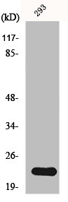
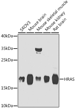
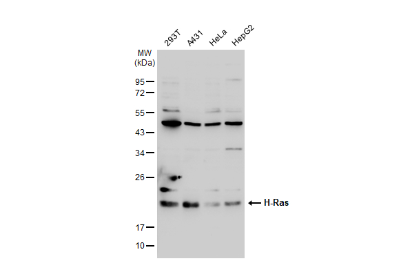
- Application Supplier NoteThe binding reactivity of this antibody to H-p21Ras, K-p21Ras and N-p21Ras protein was determined using ELISA. This antibody was also capable of recognizing wild type and mutated ras protein from various normal and tumor cell lines using western blot and immunocytometry assay. This antibody presented strong immunoreactivity for 14 primary human cancer tissues but showed weak or negative immunoreactivity in the corresponding normal tissues. It was reported to bind positively with QGY7703, SMMC7721, HepG2: hepatocarcinoma; BGC853, MKN28: gastric cancer; HCT116: colorectal cancer; T24: bladder cancer; SKOV3: ovary cancer; MDA-MB-231, MDA-MB-435, MCF7: breast cancer; HeLa: cervical cancer; Hep2: laryngocarcinoma; C8166, K562 cell lines (PMID: 26897358). This antibody was also used in the immunohistochemical staining of FFPE section of colorectal tissues, colorectal inflammatory polyps, colorectal low-grade intraepithelial neoplasia, colorectal high-grade intraepithelial neoplasia, invasive colorectal carcinomas and corresponding adjacent tissues. The immunoreactivity was measured by evaluating percentage of positive cells and histological score (HSCORE) (PMID: 21138685). This antibody can also be used in the immunofluorescent staining of various normal and cancer tissues (PMID: 32664770).
- ApplicationsImmunoFluorescence, Western Blot, ELISA, ImmunoCytoChemistry, ImmunoHistoChemistry, ImmunoHistoChemistry Paraffin
- CertificationResearch Use Only
- ClonalityMonoclonal
- Clone IDKGH-R1
- Gene ID3265
- Target nameHRAS
- Target descriptionHRas proto-oncogene, GTPase
- Target synonymsC-BAS/HAS, C-H-RAS, C-HA-RAS1, CTLO, H-RASIDX, HAMSV, HRAS1, RASH1, p21ras, GTPase HRas, GTP- and GDP-binding peptide B, Ha-Ras1 proto-oncoprotein, Harvey rat sarcoma viral oncogene homolog, Harvey rat sarcoma viral oncoprotein, Ras family small GTP binding protein H-Ras, c-has/bas p21 protein, p19 H-RasIDX protein, transformation gene: oncogene HAMSV, transforming protein p21, v-Ha-ras Harvey rat sarcoma viral oncogene homolog
- HostHuman
- IsotypeIgG1
- Protein IDP01111
- Protein NameGTPase NRas
- Scientific DescriptionThis chimeric human antibody was made using the variable domain sequences of the original Mouse IgG2b format, for improved compatibility with existing reagents, assays and techniques.
- ReactivityHuman
- Storage Instruction-20°C,2°C to 8°C
- UNSPSC41116161
Related products
Product group Antibodies
NRAS/HRAS/KRAS AntibodyCSB-PA003358
ApplicationsWestern Blot, ELISA, ImmunoHistoChemistry
ReactivityHuman, Mouse, Rat
TargetHRAS
- SizePrice

Product group Antibodies
Anti-GTPase HRAS Antibody Picoband(r)A00114-CARRIER-FREE
ApplicationsWestern Blot
ReactivityHuman, Mouse, Rat
TargetHRAS
- SizePrice
Product group Antibodies
ApplicationsWestern Blot, ImmunoHistoChemistry
ReactivityHuman, Mouse, Rat
- SizePrice
Product group Antibodies

HRAS / H-Ras AntibodyLS-C831541
ApplicationsImmunoHistoChemistry
ReactivityHuman, Mouse, Rat
TargetHRAS
- SizePrice

Product group Antibodies
Anti-HRAS AntibodyHPA070222
ApplicationsImmunoCytoChemistry
ReactivityHuman
TargetHRAS
- SizePrice
Product group Antibodies
ApplicationsImmunoPrecipitation, Western Blot, ImmunoCytoChemistry, ImmunoHistoChemistry
ReactivityMouse
TargetHRAS
- SizePrice
Product group Antibodies
HRAS Recombinant AntibodyBSM-61138R
ApplicationsFlow Cytometry, ImmunoPrecipitation
TargetHRAS
- SizePrice
Product group Antibodies
H-Ras antibodyGTX116041
ApplicationsImmunoFluorescence, ImmunoPrecipitation, Western Blot, ImmunoCytoChemistry, ImmunoHistoChemistry, ImmunoHistoChemistry Paraffin
ReactivityHuman, Mouse, Rat
TargetHRAS
- SizePrice
Product group Antibodies
Anti-HRAS Antibody144-07901
ApplicationsWestern Blot, ImmunoHistoChemistry
ReactivityHuman, Mouse, Rat
TargetHRAS
- SizePrice