


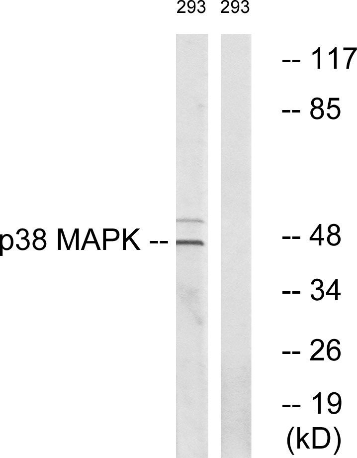
Figure 1. Western blot analysis of P38 Alpha/MAPK14 using anti-P38 Alpha/MAPK14 antibody (A00176-2). Electrophoresis was performed on a 5-20% SDS-PAGE gel at 70V (Stacking gel) / 90V (Resolving gel) for 2-3 hours. The sample well of each lane was loaded with 30 ug of sample under reducing conditions. Lane 1: human Jurkat whole cell lysates, Lane 2: human SW620 whole cell lysates, Lane 3: human Hela whole cell lysates, Lane 4: rat lung tissue lysates, Lane 5: rat heart tissue lysates, Lane 6: mouse lung tissue lysates, Lane 7: mouse heart tissue lysates. After electrophoresis, proteins were transferred to a nitrocellulose membrane at 150 mA for 50-90 minutes. Blocked the membrane with 5% non-fat milk/TBS for 1.5 hour at RT. The membrane was incubated with rabbit anti-P38 Alpha/MAPK14 antigen affinity purified polyclonal antibody (Catalog # A00176-2) at 0.5 microg/mL overnight at 4°C, then washed with TBS-0.1%Tween 3 times with 5 minutes each and probed with a goat anti-rabbit IgG-HRP secondary antibody at a dilution of 1:5000 for 1.5 hour at RT. The signal is developed using an Enhanced Chemiluminescent detection (ECL) kit (Catalog # EK1002) with Tanon 5200 system. A specific band was detected for P38 Alpha/MAPK14 at approximately 41 kDa. The expected band size for P38 Alpha/MAPK14 is at 41 kDa.
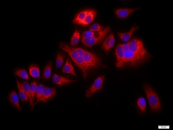
. P38 Alpha/MAPK14 was detected in an immunocytochemical section of Caco-2 cells. Enzyme antigen retrieval was performed using IHC enzyme antigen retrieval reagent (AR0022) for 15 mins. The cells were blocked with 10% goat serum. And then incubated with 5 microg/mL rabbit anti-P38 Alpha/MAPK14 Antibody (A00176-2) overnight at 4°C. DyLight®488 Conjugated Goat Anti-Rabbit IgG (BA1127) was used as secondary antibody at 1:100 dilution and incubated for 30 minutes at 37°C. The section was counterstained with DAPI. Visualize using a fluorescence microscope and filter sets appropriate for the label used.")
. Overlay histogram showing 293T cells stained with A00176-2 (Blue line). To facilitate intracellular staining, cells were fixed with 4% paraformaldehyde and permeabilized with permeabilization buffer. The cells were blocked with 10% normal goat serum. And then incubated with rabbit anti-P38 Alpha/MAPK14 Antibody (A00176-2, 1 microg/1x106 cells) for 30 min at 20°C. DyLight®488 conjugated goat anti-rabbit IgG (BA1127, 5-10 microg/1x106 cells) was used as secondary antibody for 30 minutes at 20°C. Isotype control antibody (Green line) was rabbit IgG (1 microg/1x106) used under the same conditions. Unlabelled sample without incubation with primary antibody and secondary antibody (Red line) was used as a blank control.")
Figure 1. Western blot analysis of P38 Alpha/MAPK14 using anti-P38 Alpha/MAPK14 antibody (A00176-2). Electrophoresis was performed on a 5-20% SDS-PAGE gel at 70V (Stacking gel) / 90V (Resolving gel) for 2-3 hours. The sample well of each lane was loaded with 30 ug of sample under reducing conditions. Lane 1: human Jurkat whole cell lysates, Lane 2: human SW620 whole cell lysates, Lane 3: human Hela whole cell lysates, Lane 4: rat lung tissue lysates, Lane 5: rat heart tissue lysates, Lane 6: mouse lung tissue lysates, Lane 7: mouse heart tissue lysates. After electrophoresis, proteins were transferred to a nitrocellulose membrane at 150 mA for 50-90 minutes. Blocked the membrane with 5% non-fat milk/TBS for 1.5 hour at RT. The membrane was incubated with rabbit anti-P38 Alpha/MAPK14 antigen affinity purified polyclonal antibody (Catalog # A00176-2) at 0.5 microg/mL overnight at 4°C, then washed with TBS-0.1%Tween 3 times with 5 minutes each and probed with a goat anti-rabbit IgG-HRP secondary antibody at a dilution of 1:5000 for 1.5 hour at RT. The signal is developed using an Enhanced Chemiluminescent detection (ECL) kit (Catalog # EK1002) with Tanon 5200 system. A specific band was detected for P38 Alpha/MAPK14 at approximately 41 kDa. The expected band size for P38 Alpha/MAPK14 is at 41 kDa.
Anti-p38 alpha/MAPK14 Antibody Picoband(r)
A00176-2-CARRIER-FREE
ApplicationsFlow Cytometry, ImmunoPrecipitation, Western Blot, ImmunoHistoChemistry
Product group Antibodies
ReactivityHuman, Mouse, Rat
TargetMAPK14
Overview
- SupplierBoster Bio
- Product NameAnti-p38 alpha/MAPK14 Antibody Picoband(r)
- Delivery Days Customer9
- ApplicationsFlow Cytometry, ImmunoPrecipitation, Western Blot, ImmunoHistoChemistry
- CertificationResearch Use Only
- ClonalityPolyclonal
- Concentration500 ug/ml
- Gene ID1432
- Target nameMAPK14
- Target descriptionmitogen-activated protein kinase 14
- Target synonymsCSBP, CSBP1, CSBP2, CSPB1, EXIP, Mxi2, PRKM14, PRKM15, RK, SAPK2A, p38, p38ALPHA, mitogen-activated protein kinase 14, CSAID-binding protein, MAP kinase 14, MAP kinase Mxi2, MAP kinase p38 alpha, MAX-interacting protein 2, cytokine suppressive anti-inflammatory drug binding protein, mitogen-activated protein kinase p38 alpha, p38 MAP kinase, p38 mitogen activated protein kinase, p38alpha Exip, stress-activated protein kinase 2A
- HostRabbit
- IsotypeIgG
- Protein IDQ16539
- Protein NameMitogen-activated protein kinase 14
- Scientific DescriptionBoster Bio Anti-p38 alpha/MAPK14 Antibody Picoband® catalog # A00176-2. Tested in Flow Cytometry, IP, IHC, WB applications. This antibody reacts with Human, Mouse, Rat. The brand Picoband indicates this is a premium antibody that guarantees superior quality, high affinity, and strong signals with minimal background in Western blot applications. Only our best-performing antibodies are designated as Picoband, ensuring unmatched performance.
- ReactivityHuman, Mouse, Rat
- Storage Instruction-20°C,2°C to 8°C
- UNSPSC12352203
Related products
Product group Antibodies
Anti-p38 MAPK AntibodyA94875
ApplicationsImmunoFluorescence, Western Blot, ELISA, ImmunoHistoChemistry
ReactivityHuman, Mouse, Rat
- SizePrice
Product group Antibodies
References
P38 MAPK Polyclonal AntibodyBS-0637R
ApplicationsFlow Cytometry, ImmunoFluorescence, Western Blot, ELISA, ImmunoCytoChemistry, ImmunoHistoChemistry, ImmunoHistoChemistry Frozen, ImmunoHistoChemistry Paraffin
ReactivityCanine, Human, Mouse, Rabbit, Rat, Sheep
TargetMAPK14
- SizePrice
Product group Antibodies
MAPK14 Monoclonal AntibodyCSB-MA135943
ApplicationsELISA, ImmunoHistoChemistry
ReactivityHuman, Mouse, Rat
TargetMAPK14
- SizePrice
Product group Antibodies
MAPK14 Polyclonal AntibodyCAC14688
ApplicationsWestern Blot, ELISA, ImmunoHistoChemistry
TargetMAPK14
- SizePrice
Product group Antibodies

ApplicationsELISA
ReactivityHuman
TargetMAPK14
- SizePrice
Product group Antibodies
p38 MAPK antibody [N1C3-2]GTX110720
ApplicationsImmunoFluorescence, Western Blot, ImmunoCytoChemistry, ImmunoHistoChemistry, ImmunoHistoChemistry Paraffin
ReactivityCanine, Human, Mouse, Rat
TargetMAPK14
- SizePrice
Product group Antibodies
Anti-p38 MAPK AntibodyCAB0227
ApplicationsWestern Blot, ELISA, ImmunoHistoChemistry, ImmunoHistoChemistry Paraffin
ReactivityHuman
TargetMAPK14
- SizePrice
Product group Antibodies
ApplicationsWestern Blot, ImmunoHistoChemistry
ReactivityHuman
TargetMAPK14
- SizePrice