

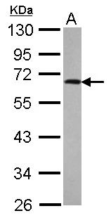
anti-p70S6K (human), Rabbit Monoclonal (RM438)
REV-31-1329-00
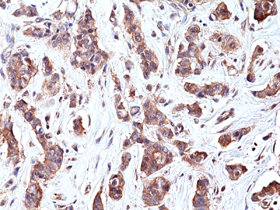
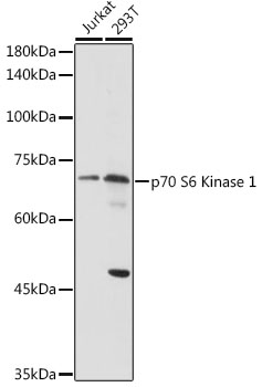
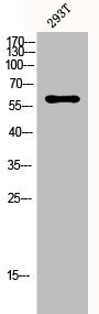
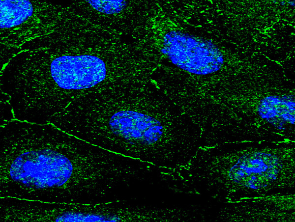
ApplicationsWestern Blot, ImmunoHistoChemistry
Product group Antibodies
ReactivityHuman
TargetRPS6KB1
Overview
- SupplierRevMAb Biosciences
- Product Nameanti-p70S6K (human), Rabbit Monoclonal (RM438)
- Delivery Days Customer2
- ApplicationsWestern Blot, ImmunoHistoChemistry
- CertificationResearch Use Only
- ClonalityMonoclonal
- Clone IDRM438
- Gene ID6198
- Target nameRPS6KB1
- Target descriptionribosomal protein S6 kinase B1
- Target synonymsPS6K, S6K, S6K-beta-1, S6K1, STK14A, p70 S6KA, p70(S6K)-alpha, p70-S6K, p70-alpha, ribosomal protein S6 kinase beta-1, ribosomal protein S6 kinase I, ribosomal protein S6 kinase, 70kDa, polypeptide 1, serine/threonine kinase 14 alpha, serine/threonine-protein kinase 14A
- HostRabbit
- IsotypeIgG
- Protein IDP23443
- Protein NameRibosomal protein S6 kinase beta-1
- Scientific DescriptionRecombinant Antibody. This antibody reacts to human p70S6K. Apllication: IHC, WB. Liquid. 50% Glycerol/PBS with 1% BSA and 0.09% sodium azide. The p70S6 kinase (p70 Ribosomal Protein S6 Kinase, p70S6K) is a 70kDa member of the ribosomal S6 kinase (RSK) family of serine/threonine kinases. p70S6 kinase is predominantly localized in the cytoplasm and is essential in growth factors regulated cell proliferation, pathways involving cell motility, such as metastases, the immune response and tissue repair. p70S6 kinase acts downstream of phosphoinositide (PI) 3-kinase and its main physiological target is the S6 ribosomal protein, which is involved in upregulation of protein synthesis. The kinase activity of p70S6 kinase leads to an increase in protein synthesis and cell proliferation. Amplification of the region of DNA encoding p70S6 kinase and overexpression are seen in some breast cancer cell lines. - The p70S6 kinase (p70 Ribosomal Protein S6 Kinase, p70S6K) is a 70kDa member of the ribosomal S6 kinase (RSK) family of serine/threonine kinases. p70S6 kinase is predominantly localized in the cytoplasm and is essential in growth factors regulated cell proliferation, pathways involving cell motility, such as metastases, the immune response and tissue repair. p70S6 kinase acts downstream of phosphoinositide (PI) 3-kinase and its main physiological target is the S6 ribosomal protein, which is involved in upregulation of protein synthesis. The kinase activity of p70S6 kinase leads to an increase in protein synthesis and cell proliferation. Amplification of the region of DNA encoding p70S6 kinase and overexpression are seen in some breast cancer cell lines.
- ReactivityHuman
- Storage Instruction-20°C,2°C to 8°C
- UNSPSC41116161
Datasheet
Related products
Product group Antibodies
Anti-RPS6KB1 Antibody144-02190
ApplicationsWestern Blot
ReactivityHuman, Mouse
TargetRPS6KB1
- SizePrice
Product group Antibodies
Anti-S6K1 AntibodyA13944
ApplicationsImmunoFluorescence, Western Blot, ImmunoCytoChemistry, ImmunoHistoChemistry
ReactivityHuman, Mouse, Rat
- SizePrice
Product group Antibodies

ApplicationsImmunoHistoChemistry, ImmunoHistoChemistry Paraffin
ReactivityHuman, Mouse, Rat
TargetRPS6KB1
- SizePrice

Product group Antibodies
Anti-S6K1/RPS6KB1 Antibody Picoband(r)A01475-2-CARRIER-FREE
ApplicationsFlow Cytometry, Western Blot
ReactivityHuman, Mouse
TargetRPS6KB1
- SizePrice
Product group Antibodies
RPS6KB1 AntibodyCSB-PA003686
ApplicationsImmunoFluorescence, Western Blot, ELISA, ImmunoHistoChemistry
ReactivityHuman, Mouse, Rat
TargetRPS6KB1
- SizePrice
Product group Antibodies

Goat anti-p70S6K / RPS6KB1EB05630
ApplicationsWestern Blot, ELISA
ReactivityBovine, Canine, Human, Mouse, Rat
TargetRPS6KB1
- SizePrice
Product group Antibodies
Rps6Kb1 Recombinant AntibodyCAC12413
ApplicationsImmunoFluorescence, ELISA
TargetRPS6KB1
- SizePrice
Product group Antibodies
RPS6KB1 Polyclonal AntibodyBS-1426R
ApplicationsFlow Cytometry, ImmunoFluorescence, Western Blot, ELISA, ImmunoCytoChemistry, ImmunoHistoChemistry, ImmunoHistoChemistry Frozen, ImmunoHistoChemistry Paraffin
ReactivityBovine, Canine, Chicken, Human, Mouse, Porcine, Rat
TargetRPS6KB1
- SizePrice

Product group Antibodies
Anti-RPS6KB1 AntibodyHPA039442
ApplicationsWestern Blot, ImmunoCytoChemistry, ImmunoHistoChemistry
ReactivityHuman
TargetRPS6KB1
- SizePrice
Product group Antibodies
p70 S6K antibodyGTX107562
ApplicationsImmunoFluorescence, Western Blot, ImmunoCytoChemistry, ImmunoHistoChemistry, ImmunoHistoChemistry Paraffin
ReactivityHuman, Mouse, Rat
TargetRPS6KB1
- SizePrice