


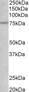
Figure 1. Western blot analysis of p75 NGF Receptor/NGFR using anti-p75 NGF Receptor/NGFR antibody (A01187). Electrophoresis was performed on a 5-20% SDS-PAGE gel at 70V (Stacking gel) / 90V (Resolving gel) for 2-3 hours. The sample well of each lane was loaded with 30ug of sample under reducing conditions. Lane 1: rat brain tissue lysates, Lane 2: human Sw620 whole cell lysates, Lane 3: human Caco-2 whole cell lysates. After Electrophoresis, proteins were transferred to a Nitrocellulose membrane at 150mA for 50-90 minutes. Blocked the membrane with 5% Non-fat Milk/ TBS for 1.5 hour at RT. The membrane was incubated with rabbit anti-p75 NGF Receptor/NGFR antigen affinity purified polyclonal antibody (Catalog # A01187) at 0.5 microg/mL overnight at 4°C, then washed with TBS-0.1%Tween 3 times with 5 minutes each and probed with a goat anti-rabbit IgG-HRP secondary antibody at a dilution of 1:5000 for 1.5 hour at RT. The signal is developed using an Enhanced Chemiluminescent detection (ECL) kit (Catalog # EK1002) with Tanon 5200 system. A specific band was detected for p75 NGF Receptor/NGFR at approximately 45KD,75KD. The expected band size for p75 NGF Receptor/NGFR is at 45KD,75KD.
. p75 NGF Receptor/NGFR was detected in immunocytochemical section of A431 cells. Enzyme antigen retrieval was performed using IHC enzyme antigen retrieval reagent (AR0022) for 15 mins. The cells were blocked with 10% goat serum. And then incubated with 2microg/mL rabbit anti-p75 NGF Receptor/NGFR Antibody (A01187) overnight at 4°C. DyLight®488 Conjugated Goat Anti-Rabbit IgG (BA1127) was used as secondary antibody at 1:100 dilution and incubated for 30 minutes at 37°C. The section was counterstained with DAPI. Visualize using a fluorescence microscope and filter sets appropriate for the label used.")
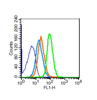
. Overlay histogram showing A431 cells stained with A01187 (Blue line). To facilitate intracellular staining, cells were fixed with 4% paraformaldehyde and permeabilized with permeabilization buffer. The cells were blocked with 10% normal goat serum. And then incubated with rabbit anti-p75 NGF Receptor/NGFR Antibody (A01187, 1microg/1x106 cells) for 30 min at 20°C. DyLight®488 conjugated goat anti-rabbit IgG (BA1127, 5-10microg/1x106 cells) was used as secondary antibody for 30 minutes at 20°C. Isotype control antibody (Green line) was rabbit IgG (1microg/1x106) used under the same conditions. Unlabelled sample without incubation with primary antibody and secondary antibody (Red line) was used as a blank control.")
Figure 1. Western blot analysis of p75 NGF Receptor/NGFR using anti-p75 NGF Receptor/NGFR antibody (A01187). Electrophoresis was performed on a 5-20% SDS-PAGE gel at 70V (Stacking gel) / 90V (Resolving gel) for 2-3 hours. The sample well of each lane was loaded with 30ug of sample under reducing conditions. Lane 1: rat brain tissue lysates, Lane 2: human Sw620 whole cell lysates, Lane 3: human Caco-2 whole cell lysates. After Electrophoresis, proteins were transferred to a Nitrocellulose membrane at 150mA for 50-90 minutes. Blocked the membrane with 5% Non-fat Milk/ TBS for 1.5 hour at RT. The membrane was incubated with rabbit anti-p75 NGF Receptor/NGFR antigen affinity purified polyclonal antibody (Catalog # A01187) at 0.5 microg/mL overnight at 4°C, then washed with TBS-0.1%Tween 3 times with 5 minutes each and probed with a goat anti-rabbit IgG-HRP secondary antibody at a dilution of 1:5000 for 1.5 hour at RT. The signal is developed using an Enhanced Chemiluminescent detection (ECL) kit (Catalog # EK1002) with Tanon 5200 system. A specific band was detected for p75 NGF Receptor/NGFR at approximately 45KD,75KD. The expected band size for p75 NGF Receptor/NGFR is at 45KD,75KD.
Anti-p75 NGF Receptor/NGFR Antibody Picoband(r)
A01187-CARRIER-FREE
ApplicationsFlow Cytometry, ImmunoFluorescence, Western Blot, ELISA, ImmunoCytoChemistry
Product group Antibodies
ReactivityHuman, Rat
TargetNGFR
Overview
- SupplierBoster Bio
- Product NameAnti-p75 NGF Receptor/NGFR Antibody Picoband(r)
- Delivery Days Customer9
- Application Supplier NoteTested Species: In-house tested species with positive results. Other applications have not been tested. Optimal dilutions should be determined by end users.
- ApplicationsFlow Cytometry, ImmunoFluorescence, Western Blot, ELISA, ImmunoCytoChemistry
- CertificationResearch Use Only
- ClonalityPolyclonal
- Concentration500 ug/ml
- Gene ID4804
- Target nameNGFR
- Target descriptionnerve growth factor receptor
- Target synonymsCD271, Gp80-LNGFR, TNFRSF16, p75(NTR), p75NTR, tumor necrosis factor receptor superfamily member 16, NGF receptor, TNFR superfamily, member 16, low affinity neurotrophin receptor p75NTR, low-affinity nerve growth factor receptor, low-affinity nerve growth factor receptor p75NGFR, low-affinity nerve growth factor receptor p75NGR, p75 ICD
- HostRabbit
- IsotypeIgG
- Protein IDP08138
- Protein NameTumor necrosis factor receptor superfamily member 16
- Scientific DescriptionBoster Bio Anti-p75 NGF Receptor/NGFR Antibody Picoband® catalog # A01187. Tested in ELISA, Flow Cytometry, IF, ICC, WB applications. This antibody reacts with Human, Rat. The brand Picoband indicates this is a premium antibody that guarantees superior quality, high affinity, and strong signals with minimal background in Western blot applications. Only our best-performing antibodies are designated as Picoband, ensuring unmatched performance.
- ReactivityHuman, Rat
- Storage Instruction-20°C,2°C to 8°C
- UNSPSC12352203
Related products
Product group Antibodies
ApplicationsWestern Blot, ELISA
ReactivityHuman, Rat
- SizePrice
Product group Antibodies
Anti-NGFR Antibody144-02097
ApplicationsWestern Blot
ReactivityHuman, Mouse
TargetNGFR
- SizePrice
Product group Antibodies

NGFR / CD271 / TNR16 AntibodyLS-C746900
ApplicationsWestern Blot
ReactivityHuman, Mouse, Rat
TargetNGFR
- SizePrice
Product group Antibodies
NGFR/p75NTR Polyclonal AntibodyBS-7122R
ApplicationsFlow Cytometry, ImmunoFluorescence, Western Blot, ELISA, ImmunoCytoChemistry, ImmunoHistoChemistry, ImmunoHistoChemistry Frozen, ImmunoHistoChemistry Paraffin
ReactivityCanine, Human, Mouse, Rat
TargetNGFR
- SizePrice
Product group Antibodies
NGFR AntibodyCSB-PA003447
ApplicationsWestern Blot, ELISA
ReactivityHuman, Mouse, Rat
TargetNGFR
- SizePrice
Product group Antibodies

Goat anti-NGFREB06775
ApplicationsWestern Blot, ELISA
ReactivityBovine, Canine, Human, Mouse, Rat
TargetNGFR
- SizePrice
Product group Antibodies
NGFR Polyclonal AntibodyCAC14773
ApplicationsWestern Blot, ELISA, ImmunoHistoChemistry
ReactivityMouse
TargetNGFR
- SizePrice
![p75 NGF Receptor / CD271 antibody detects p75 NGF Receptor / CD271 protein by immunofluorescent analysis. Sample: DIV10 rat E18 primary cortical neuron cells were fixed in 4% paraformaldehyde at RT for 15 min. Green: p75 NGF Receptor / CD271 stained by p75 NGF Receptor / CD271 antibody (GTX102262) diluted at 1:500. Red: Tau, stained by Tau antibody [GT287] (GTX634809) diluted at 1:500. Blue: Fluoroshield with DAPI (GTX30920).](https://www.genetex.com/upload/website/prouct_img/normal/GTX102262/GTX102262_43811_20200820_ICC_IF_R_w_23060100_660.webp)
Product group Antibodies
p75 NGF Receptor / CD271 antibodyGTX102262
ApplicationsFlow Cytometry, ImmunoFluorescence, Western Blot, ImmunoCytoChemistry, ImmunoHistoChemistry, ImmunoHistoChemistry Frozen, ImmunoHistoChemistry Paraffin
ReactivityHuman, Mouse, Rat
TargetNGFR
- SizePrice

Product group Antibodies
Anti-NGFR AntibodyHPA004765
ApplicationsWestern Blot, ImmunoCytoChemistry, ImmunoHistoChemistry
ReactivityHuman
TargetNGFR
- SizePrice