

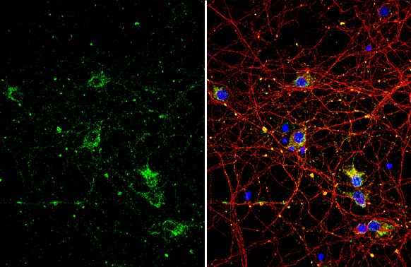
p75 NGF Receptor / CD271 antibody detects p75 NGF Receptor / CD271 protein by immunofluorescent analysis. Sample: DIV10 rat E18 primary cortical neuron cells were fixed in 4% paraformaldehyde at RT for 15 min. Green: p75 NGF Receptor / CD271 stained by p75 NGF Receptor / CD271 antibody (GTX102262) diluted at 1:500. Red: Tau, stained by Tau antibody [GT287] (GTX634809) diluted at 1:500. Blue: Fluoroshield with DAPI (GTX30920).
 diluted at 1:500.")
![p75 NGF Receptor antibody detects p75 NGF Receptor protein expression by immunohistochemical analysis. Sample: Frozen sectioned E13.5 Rat brain. Green: p75 NGF Receptor protein stained by p75 NGF Receptor antibody (GTX102262) diluted at 1:250. Red: beta Tubulin 3/ TUJ1, a mature neuron marker, stained by beta Tubulin 3/ TUJ1 antibody [GT11710] (GTX631836) diluted at 1:500. Blue: Fluoroshield with DAPI (GTX30920).](https://www.genetex.com/upload/website/prouct_img/normal/GTX102262/GTX102262_40730_20160921_IHC-Fr_w_23060100_564.webp "p75 NGF Receptor antibody detects p75 NGF Receptor protein expression by immunohistochemical analysis. Sample: Frozen sectioned E13.5 Rat brain. Green: p75 NGF Receptor protein stained by p75 NGF Receptor antibody (GTX102262) diluted at 1:250. Red: beta Tubulin 3/ TUJ1, a mature neuron marker, stained by beta Tubulin 3/ TUJ1 antibody [GT11710] (GTX631836) diluted at 1:500. Blue: Fluoroshield with DAPI (GTX30920).")
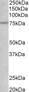
 and treated (+) PC-3 whole cell extracts (30 μg) were separated by 10% SDS-PAGE, and the membrane was blotted with p75 NGF Receptor / CD271 antibody (GTX102262) diluted at 1:1000. The HRP-conjugated anti-rabbit IgG antibody (GTX213110-01) was used to detect the primary antibody.")
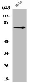
 was separated by 10% SDS-PAGE, and the membrane was blotted with p75 NGF Receptor antibody (GTX102262) at a dilution of 1:2000.")
 were separated by 10% SDS-PAGE, and the membrane was blotted with p75 NGF Receptor / CD271 antibody (GTX102262) diluted at 1:2000. The HRP-conjugated anti-rabbit IgG antibody (GTX213110-01) was used to detect the primary antibody.")
p75 NGF Receptor / CD271 antibody detects p75 NGF Receptor / CD271 protein by immunofluorescent analysis. Sample: DIV10 rat E18 primary cortical neuron cells were fixed in 4% paraformaldehyde at RT for 15 min. Green: p75 NGF Receptor / CD271 stained by p75 NGF Receptor / CD271 antibody (GTX102262) diluted at 1:500. Red: Tau, stained by Tau antibody [GT287] (GTX634809) diluted at 1:500. Blue: Fluoroshield with DAPI (GTX30920).
p75 NGF Receptor / CD271 antibody
GTX102262
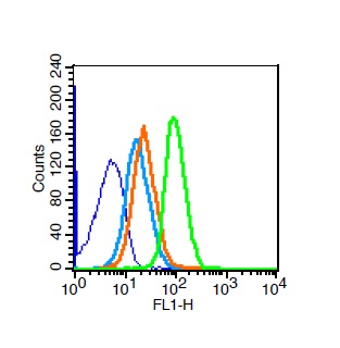
ApplicationsFlow Cytometry, ImmunoFluorescence, Western Blot, ImmunoCytoChemistry, ImmunoHistoChemistry, ImmunoHistoChemistry Frozen, ImmunoHistoChemistry Paraffin
Product group Antibodies
ReactivityHuman, Mouse, Rat
TargetNGFR
Overview
- SupplierGeneTex
- Product Namep75 NGF Receptor / CD271 antibody
- Delivery Days Customer9
- Application Supplier NoteWB: 1:500-1:3000. ICC/IF: 1:100-1:1000. IHC-P: 1:100-1:1000. IHC-Fr: 1:100-1:1000. FCM: Assay dependent. *Optimal dilutions/concentrations should be determined by the researcher.Not tested in other applications.
- ApplicationsFlow Cytometry, ImmunoFluorescence, Western Blot, ImmunoCytoChemistry, ImmunoHistoChemistry, ImmunoHistoChemistry Frozen, ImmunoHistoChemistry Paraffin
- CertificationResearch Use Only
- ClonalityPolyclonal
- Concentration0.43 mg/ml
- ConjugateUnconjugated
- Gene ID4804
- Target nameNGFR
- Target descriptionnerve growth factor receptor
- Target synonymsCD271, Gp80-LNGFR, TNFRSF16, p75(NTR), p75NTR, tumor necrosis factor receptor superfamily member 16, NGF receptor, TNFR superfamily, member 16, low affinity neurotrophin receptor p75NTR, low-affinity nerve growth factor receptor, low-affinity nerve growth factor receptor p75NGFR, low-affinity nerve growth factor receptor p75NGR, p75 ICD
- HostRabbit
- IsotypeIgG
- Protein IDP08138
- Protein NameTumor necrosis factor receptor superfamily member 16
- Scientific DescriptionNerve growth factor receptor contains an extracellular domain containing four 40-amino acid repeats with 6 cysteine residues at conserved positions followed by a serine/threonine-rich region, a single transmembrane domain, and a 155-amino acid cytoplasmic domain. The cysteine-rich region contains the nerve growth factor binding domain. [provided by RefSeq]
- ReactivityHuman, Mouse, Rat
- Storage Instruction-20°C or -80°C,2°C to 8°C
- UNSPSC41116161
Datasheet
Related products
Product group Antibodies
ApplicationsWestern Blot, ELISA
ReactivityHuman, Rat
- SizePrice

Product group Antibodies
Anti-p75 NGF Receptor/NGFR Antibody Picoband(r)A01187-CARRIER-FREE
ApplicationsFlow Cytometry, ImmunoFluorescence, Western Blot, ELISA, ImmunoCytoChemistry
ReactivityHuman, Rat
TargetNGFR
- SizePrice
Product group Antibodies
Anti-NGFR Antibody144-02097
ApplicationsWestern Blot
ReactivityHuman, Mouse
TargetNGFR
- SizePrice
Product group Antibodies

NGFR / CD271 / TNR16 AntibodyLS-C746900
ApplicationsWestern Blot
ReactivityHuman, Mouse, Rat
TargetNGFR
- SizePrice
Product group Antibodies
NGFR/p75NTR Polyclonal AntibodyBS-7122R
ApplicationsFlow Cytometry, ImmunoFluorescence, Western Blot, ELISA, ImmunoCytoChemistry, ImmunoHistoChemistry, ImmunoHistoChemistry Frozen, ImmunoHistoChemistry Paraffin
ReactivityCanine, Human, Mouse, Rat
TargetNGFR
- SizePrice
Product group Antibodies
NGFR AntibodyCSB-PA003447
ApplicationsWestern Blot, ELISA
ReactivityHuman, Mouse, Rat
TargetNGFR
- SizePrice
Product group Antibodies

Goat anti-NGFREB06775
ApplicationsWestern Blot, ELISA
ReactivityBovine, Canine, Human, Mouse, Rat
TargetNGFR
- SizePrice
Product group Antibodies
NGFR Polyclonal AntibodyCAC14773
ApplicationsWestern Blot, ELISA, ImmunoHistoChemistry
ReactivityMouse
TargetNGFR
- SizePrice

Product group Antibodies
ApplicationsImmunoHistoChemistry, ImmunoHistoChemistry Paraffin, Other Application
ReactivityHuman, Primate
TargetNGFR
- SizePrice
![IHC-P analysis of human breast carcinoma tissue using GTX17979 p75 NGF Receptor / CD271 antibody [NGFR/1964].](https://www.genetex.com/upload/website/prouct_img/normal/GTX17979/GTX17979_20200115_IHC-P_393_w_23060620_367.webp)
Product group Antibodies
ApplicationsImmunoHistoChemistry, ImmunoHistoChemistry Paraffin
ReactivityHuman
TargetNGFR
- SizePrice