


Immunohistochemical staining of human testis shows strong cytoplasmic positivity in cells in seminiferous ducts.

Immunohistochemical staining of human testis shows strong cytoplasmic positivity in cells in seminiferous ducts.
Anti-PAIP1 Antibody
HPA073653
ApplicationsWestern Blot, ImmunoHistoChemistry
Product group Antibodies
ReactivityHuman
TargetPAIP1
Overview
- SupplierAtlas Antibodies
- Product NameAnti-PAIP1 Antibody
- Delivery Days Customer4
- ApplicationsWestern Blot, ImmunoHistoChemistry
- CertificationResearch Use Only
- ClonalityPolyclonal
- ConjugateUnconjugated
- Gene ID10605
- Target namePAIP1
- Target descriptionpoly(A) binding protein interacting protein 1
- Target synonymspolyadenylate-binding protein-interacting protein 1, PABC1-interacting protein 1, PABP-interacting protein 1, PAIP-1
- HostRabbit
- IsotypeIgG
- Protein IDQ9H074
- Protein NamePolyadenylate-binding protein-interacting protein 1
- Scientific DescriptionRecombinant Protein Epitope Signature Tag (PrEST) antigen sequence
- ReactivityHuman
- Storage Instruction-20°C,2°C to 8°C
- UNSPSC41116161
Datasheet
MSDS
Related products

Product group Antibodies
Anti-PAIP1 Antibody Picoband(r)A07792-2-CARRIER-FREE
ApplicationsFlow Cytometry, ImmunoFluorescence, Western Blot, ELISA, ImmunoCytoChemistry, ImmunoHistoChemistry
ReactivityHuman
TargetPAIP1
- SizePrice
Product group Antibodies
Anti-PAIP1 AntibodyA15067
ApplicationsWestern Blot
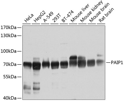
ReactivityHuman, Mouse, Rat
- SizePrice
Product group Antibodies

PAIP1 AntibodyLS-C831591
ApplicationsImmunoHistoChemistry
ReactivityHuman, Mouse, Rat
TargetPAIP1
- SizePrice

Product group Antibodies
Anti-PAIP1 AntibodyHPA076187
ApplicationsImmunoCytoChemistry
ReactivityHuman
TargetPAIP1
- SizePrice
Product group Antibodies
PAIP1 AntibodyCSB-PA880930ESR2HU
ApplicationsWestern Blot, ELISA
ReactivityHuman
TargetPAIP1
- SizePrice

Product group Antibodies
PAIP1 antibody [N3C3]GTX115446
ApplicationsWestern Blot
ReactivityHuman
TargetPAIP1
- SizePrice
Product group Antibodies
Anti-PAIP1 Antibody144-06042
ApplicationsWestern Blot
ReactivityHuman, Mouse, Rat
TargetPAIP1
- SizePrice