


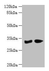
Figure 1. Western blot analysis of PBLD using anti-PBLD antibody (A10149-2). Electrophoresis was performed on a 5-20% SDS-PAGE gel at 70V (Stacking gel) / 90V (Resolving gel) for 2-3 hours. The sample well of each lane was loaded with 30 ug of sample under reducing conditions. Lane 1: human hepatocellular carcinoma tumor tissue (HCCT) lysates, Lane 2: human hepatocellular carcinoma paracancerous tissue (HCCP) lysates. Lane 3: rat liver tissue lysates, Lane 4: rat RH35 whole cell lysates, Lane 5: mouse liver tissue lysates. After electrophoresis, proteins were transferred to a nitrocellulose membrane at 150 mA for 50-90 minutes. Blocked the membrane with 5% non-fat milk/TBS for 1.5 hour at RT. The membrane was incubated with rabbit anti-PBLD antigen affinity purified polyclonal antibody (Catalog # A10149-2) at 0.5 microg/mL overnight at 4°C, then washed with TBS-0.1%Tween 3 times with 5 minutes each and probed with a goat anti-rabbit IgG-HRP secondary antibody at a dilution of 1:5000 for 1.5 hour at RT. The signal is developed using an Enhanced Chemiluminescent detection (ECL) kit (Catalog # EK1002) with Tanon 5200 system. A specific band was detected for PBLD at approximately 32 kDa. The expected band size for PBLD is at 32 kDa.
. Overlay histogram showing SiHa cells stained with A10149-2 (Blue line). To facilitate intracellular staining, cells were fixed with 4% paraformaldehyde and permeabilized with permeabilization buffer. The cells were blocked with 10% normal goat serum. And then incubated with rabbit anti-PBLD Antibody (A10149-2, 1 microg/1x106 cells) for 30 min at 20°C. DyLight®488 conjugated goat anti-rabbit IgG (BA1127, 5-10 microg/1x106 cells) was used as secondary antibody for 30 minutes at 20°C. Isotype control antibody (Green line) was rabbit IgG (1 microg/1x106) used under the same conditions. Unlabelled sample (Red line) was also used as a control.")
Figure 1. Western blot analysis of PBLD using anti-PBLD antibody (A10149-2). Electrophoresis was performed on a 5-20% SDS-PAGE gel at 70V (Stacking gel) / 90V (Resolving gel) for 2-3 hours. The sample well of each lane was loaded with 30 ug of sample under reducing conditions. Lane 1: human hepatocellular carcinoma tumor tissue (HCCT) lysates, Lane 2: human hepatocellular carcinoma paracancerous tissue (HCCP) lysates. Lane 3: rat liver tissue lysates, Lane 4: rat RH35 whole cell lysates, Lane 5: mouse liver tissue lysates. After electrophoresis, proteins were transferred to a nitrocellulose membrane at 150 mA for 50-90 minutes. Blocked the membrane with 5% non-fat milk/TBS for 1.5 hour at RT. The membrane was incubated with rabbit anti-PBLD antigen affinity purified polyclonal antibody (Catalog # A10149-2) at 0.5 microg/mL overnight at 4°C, then washed with TBS-0.1%Tween 3 times with 5 minutes each and probed with a goat anti-rabbit IgG-HRP secondary antibody at a dilution of 1:5000 for 1.5 hour at RT. The signal is developed using an Enhanced Chemiluminescent detection (ECL) kit (Catalog # EK1002) with Tanon 5200 system. A specific band was detected for PBLD at approximately 32 kDa. The expected band size for PBLD is at 32 kDa.
Anti-PBLD Antibody Picoband(r)
A10149-2-CARRIER-FREE
ApplicationsFlow Cytometry, Western Blot, ELISA
Product group Antibodies
ReactivityHuman, Mouse, Rat
TargetPBLD
Overview
- SupplierBoster Bio
- Product NameAnti-PBLD Antibody Picoband(r)
- Delivery Days Customer9
- ApplicationsFlow Cytometry, Western Blot, ELISA
- CertificationResearch Use Only
- ClonalityPolyclonal
- Concentration500 ug/ml
- Gene ID64081
- Target namePBLD
- Target descriptionphenazine biosynthesis like protein domain containing
- Target synonymsHEL-S-306, MAWBP, MAWDBP, phenazine biosynthesis-like domain-containing protein, MAWD-binding protein, epididymis secretory protein Li 306, epididymis secretory sperm binding protein
- HostRabbit
- IsotypeIgG
- Protein IDP30039
- Protein NamePhenazine biosynthesis-like domain-containing protein
- Scientific DescriptionBoster Bio Anti-PBLD Antibody Picoband® catalog # A10149-2. Tested in ELISA, WB, Flow Cytometry applications. This antibody reacts with Human, Mouse, Rat. The brand Picoband indicates this is a premium antibody that guarantees superior quality, high affinity, and strong signals with minimal background in Western blot applications. Only our best-performing antibodies are designated as Picoband, ensuring unmatched performance.
- ReactivityHuman, Mouse, Rat
- Storage Instruction-20°C,2°C to 8°C
- UNSPSC12352203
Related products
Product group Antibodies
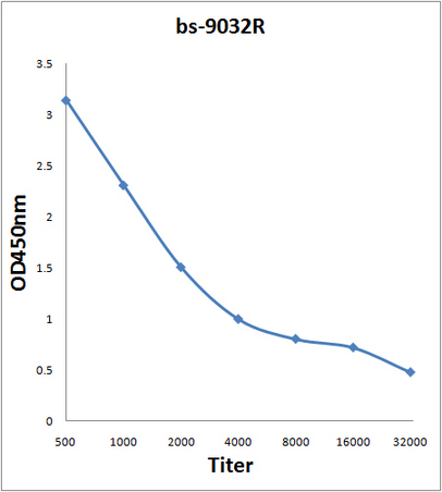
PBLD Polyclonal AntibodyBS-9032R
ApplicationsImmunoFluorescence, Western Blot, ELISA, ImmunoHistoChemistry, ImmunoHistoChemistry Paraffin
ReactivityBovine, Equine, Human, Mouse, Rat
TargetPBLD
- SizePrice
Product group Antibodies
PBLD Polyclonal AntibodyCAC13857
ApplicationsWestern Blot, ELISA, ImmunoHistoChemistry
ReactivityMouse
TargetPBLD
- SizePrice
Product group Antibodies
PBLD AntibodyCSB-PA017501LA01HU
ApplicationsWestern Blot, ELISA, ImmunoHistoChemistry
ReactivityHuman, Mouse
TargetPBLD
- SizePrice
![WB analysis of HEK293T cells transfected with PBLD plasmid (Right) or empty vector (Left) for 48 hrs using GTX83938 PBLD antibody [7G5]. Loading : 5 ug per lane](https://www.genetex.com/upload/website/prouct_img/normal/GTX83938/GTX83938_4010_WB_w_23061420_661.webp)
Product group Antibodies
PBLD antibody [7G5]GTX83938
ApplicationsFlow Cytometry, ImmunoFluorescence, Western Blot, ImmunoCytoChemistry
ReactivityHuman
TargetPBLD
- SizePrice

Product group Antibodies
Anti-PBLD AntibodyHPA038036
ApplicationsWestern Blot, ImmunoHistoChemistry
ReactivityHuman
TargetPBLD
- SizePrice
Product group Antibodies

MAWDBP / PBLD Antibody (HRP)LS-C376272
ApplicationsWestern Blot, ELISA, ImmunoHistoChemistry
ReactivityHuman, Mouse
TargetPBLD
- SizePrice
Product group Antibodies
PBLD AntibodyPACO28822
ApplicationsWestern Blot, ELISA, ImmunoHistoChemistry
ReactivityHuman, Mouse
TargetPBLD
- SizePrice