


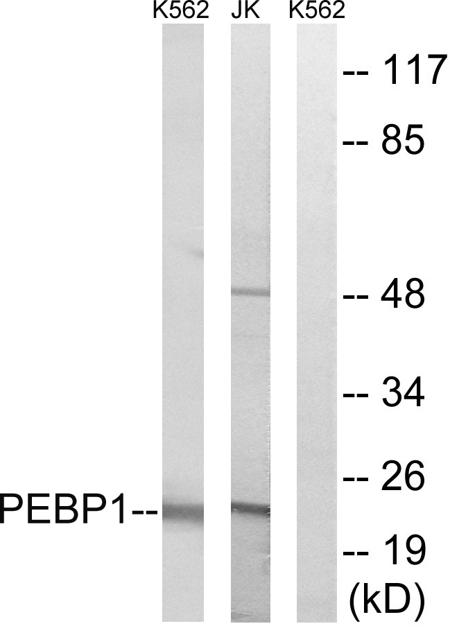
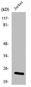
Figure 1. Western blot analysis of PBP using anti-PBP antibody (A01668). Electrophoresis was performed on a 5-20% SDS-PAGE gel at 70V (Stacking gel) / 90V (Resolving gel) for 2-3 hours. The sample well of each lane was loaded with 30 ug of sample under reducing conditions. Lane 1: human Hela whole cell lysates, Lane 2: human MCF-7 whole cell lysates, Lane 3: human HepG2 whole cell lysates, Lane 4: human 293T whole cell lysates, Lane 5: mouse testis lysates. After electrophoresis, proteins were transferred to a nitrocellulose membrane at 150 mA for 50-90 minutes. Blocked the membrane with 5% non-fat milk/TBS for 1.5 hour at RT. The membrane was incubated with rabbit anti-PBP antigen affinity purified polyclonal antibody (Catalog # A01668) at 0.5 microg/mL overnight at 4°C, then washed with TBS-0.1%Tween 3 times with 5 minutes each and probed with a goat anti-rabbit IgG-HRP secondary antibody at a dilution of 1:5000 for 1.5 hour at RT. The signal is developed using an Enhanced Chemiluminescent detection (ECL) kit (Catalog # EK1002) with Tanon 5200 system. A specific band was detected for PBP at approximately 21 kDa. The expected band size for PBP is at 21 kDa.
. PBP was detected in paraffin-embedded section of human intestinal cancer tissue. Heat mediated antigen retrieval was performed in citrate buffer (pH6, epitope retrieval solution) for 20 mins. The tissue section was blocked with 10% goat serum. The tissue section was then incubated with 1microg/ml rabbit anti-PBP Antibody (A01668) overnight at 4°C. Biotinylated goat anti-rabbit IgG was used as secondary antibody and incubated for 30 minutes at 37°C. The tissue section was developed using Strepavidin-Biotin-Complex (SABC)(Catalog # SA1022) with DAB as the chromogen.")
. PBP was detected in paraffin-embedded section of human sarcoma tissue. Heat mediated antigen retrieval was performed in citrate buffer (pH6, epitope retrieval solution) for 20 mins. The tissue section was blocked with 10% goat serum. The tissue section was then incubated with 1microg/ml rabbit anti-PBP Antibody (A01668) overnight at 4°C. Biotinylated goat anti-rabbit IgG was used as secondary antibody and incubated for 30 minutes at 37°C. The tissue section was developed using Strepavidin-Biotin-Complex (SABC)(Catalog # SA1022) with DAB as the chromogen.")
. PBP was detected in paraffin-embedded section of human endometrial carcinoma tissue. Heat mediated antigen retrieval was performed in citrate buffer (pH6, epitope retrieval solution) for 20 mins. The tissue section was blocked with 10% goat serum. The tissue section was then incubated with 1microg/ml rabbit anti-PBP Antibody (A01668) overnight at 4°C. Biotinylated goat anti-rabbit IgG was used as secondary antibody and incubated for 30 minutes at 37°C. The tissue section was developed using Strepavidin-Biotin-Complex (SABC)(Catalog # SA1022) with DAB as the chromogen.")
. PBP was detected in paraffin-embedded section of human liver cancer tissue. Heat mediated antigen retrieval was performed in citrate buffer (pH6, epitope retrieval solution) for 20 mins. The tissue section was blocked with 10% goat serum. The tissue section was then incubated with 1microg/ml rabbit anti-PBP Antibody (A01668) overnight at 4°C. Biotinylated goat anti-rabbit IgG was used as secondary antibody and incubated for 30 minutes at 37°C. The tissue section was developed using Strepavidin-Biotin-Complex (SABC)(Catalog # SA1022) with DAB as the chromogen.")
. PBP was detected in paraffin-embedded section of human mammary cancer tissue. Heat mediated antigen retrieval was performed in citrate buffer (pH6, epitope retrieval solution) for 20 mins. The tissue section was blocked with 10% goat serum. The tissue section was then incubated with 1microg/ml rabbit anti-PBP Antibody (A01668) overnight at 4°C. Biotinylated goat anti-rabbit IgG was used as secondary antibody and incubated for 30 minutes at 37°C. The tissue section was developed using Strepavidin-Biotin-Complex (SABC)(Catalog # SA1022) with DAB as the chromogen.")
. PBP was detected in paraffin-embedded section of human pancreatic cancer tissue. Heat mediated antigen retrieval was performed in citrate buffer (pH6, epitope retrieval solution) for 20 mins. The tissue section was blocked with 10% goat serum. The tissue section was then incubated with 1microg/ml rabbit anti-PBP Antibody (A01668) overnight at 4°C. Biotinylated goat anti-rabbit IgG was used as secondary antibody and incubated for 30 minutes at 37°C. The tissue section was developed using Strepavidin-Biotin-Complex (SABC)(Catalog # SA1022) with DAB as the chromogen.")
. PBP was detected in paraffin-embedded section of human tonsil tissue. Heat mediated antigen retrieval was performed in citrate buffer (pH6, epitope retrieval solution) for 20 mins. The tissue section was blocked with 10% goat serum. The tissue section was then incubated with 1microg/ml rabbit anti-PBP Antibody (A01668) overnight at 4°C. Biotinylated goat anti-rabbit IgG was used as secondary antibody and incubated for 30 minutes at 37°C. The tissue section was developed using Strepavidin-Biotin-Complex (SABC)(Catalog # SA1022) with DAB as the chromogen.")
. PBP was detected in paraffin-embedded section of mouse brain tissue. Heat mediated antigen retrieval was performed in citrate buffer (pH6, epitope retrieval solution) for 20 mins. The tissue section was blocked with 10% goat serum. The tissue section was then incubated with 1microg/ml rabbit anti-PBP Antibody (A01668) overnight at 4°C. Biotinylated goat anti-rabbit IgG was used as secondary antibody and incubated for 30 minutes at 37°C. The tissue section was developed using Strepavidin-Biotin-Complex (SABC)(Catalog # SA1022) with DAB as the chromogen.")
. PBP was detected in paraffin-embedded section of mouse kidney tissue. Heat mediated antigen retrieval was performed in citrate buffer (pH6, epitope retrieval solution) for 20 mins. The tissue section was blocked with 10% goat serum. The tissue section was then incubated with 1microg/ml rabbit anti-PBP Antibody (A01668) overnight at 4°C. Biotinylated goat anti-rabbit IgG was used as secondary antibody and incubated for 30 minutes at 37°C. The tissue section was developed using Strepavidin-Biotin-Complex (SABC)(Catalog # SA1022) with DAB as the chromogen.")
Figure 1. Western blot analysis of PBP using anti-PBP antibody (A01668). Electrophoresis was performed on a 5-20% SDS-PAGE gel at 70V (Stacking gel) / 90V (Resolving gel) for 2-3 hours. The sample well of each lane was loaded with 30 ug of sample under reducing conditions. Lane 1: human Hela whole cell lysates, Lane 2: human MCF-7 whole cell lysates, Lane 3: human HepG2 whole cell lysates, Lane 4: human 293T whole cell lysates, Lane 5: mouse testis lysates. After electrophoresis, proteins were transferred to a nitrocellulose membrane at 150 mA for 50-90 minutes. Blocked the membrane with 5% non-fat milk/TBS for 1.5 hour at RT. The membrane was incubated with rabbit anti-PBP antigen affinity purified polyclonal antibody (Catalog # A01668) at 0.5 microg/mL overnight at 4°C, then washed with TBS-0.1%Tween 3 times with 5 minutes each and probed with a goat anti-rabbit IgG-HRP secondary antibody at a dilution of 1:5000 for 1.5 hour at RT. The signal is developed using an Enhanced Chemiluminescent detection (ECL) kit (Catalog # EK1002) with Tanon 5200 system. A specific band was detected for PBP at approximately 21 kDa. The expected band size for PBP is at 21 kDa.
Anti-PBP/PEBP1 Antibody Picoband(r)
A01668-CARRIER-FREE
ApplicationsFlow Cytometry, Western Blot, ImmunoHistoChemistry
Product group Antibodies
ReactivityHuman, Mouse
TargetPEBP1
Overview
- SupplierBoster Bio
- Product NameAnti-PBP/PEBP1 Antibody Picoband(r)
- Delivery Days Customer9
- ApplicationsFlow Cytometry, Western Blot, ImmunoHistoChemistry
- CertificationResearch Use Only
- ClonalityPolyclonal
- Concentration500 ug/ml
- Gene ID5037
- Target namePEBP1
- Target descriptionphosphatidylethanolamine binding protein 1
- Target synonymsHCNP, HCNPpp, HEL-210, HEL-S-34, HEL-S-96, PBP, PEBP, PEBP-1, RKIP, phosphatidylethanolamine-binding protein 1, Raf kinase inhibitory protein, epididymis luminal protein 210, epididymis secretory protein Li 34, epididymis secretory protein Li 96, hippocampal cholinergic neurostimulating peptide, neuropolypeptide h3, prostatic binding protein, raf kinase inhibitor protein
- HostRabbit
- IsotypeIgG
- Protein IDP30086
- Protein NamePhosphatidylethanolamine-binding protein 1
- Scientific DescriptionBoster Bio Anti-PBP/PEBP1 Antibody Picoband® catalog # A01668. Tested in Flow Cytometry, IHC, WB applications. This antibody reacts with Human, Mouse. The brand Picoband indicates this is a premium antibody that guarantees superior quality, high affinity, and strong signals with minimal background in Western blot applications. Only our best-performing antibodies are designated as Picoband, ensuring unmatched performance.
- ReactivityHuman, Mouse
- Storage Instruction-20°C,2°C to 8°C
- UNSPSC12352203
Related products
Product group Antibodies
Anti-PEBP1 AntibodyA100812
ApplicationsWestern Blot, ELISA
ReactivityHuman
- SizePrice
Product group Antibodies
Anti-PEBP1 Antibody144-00578
ApplicationsImmunoFluorescence, ImmunoPrecipitation, Western Blot, Other Application
ReactivityHuman, Mouse
TargetPEBP1
- SizePrice
Product group Antibodies
PBP Recombinant AntibodyBSM-61243R
ApplicationsImmunoFluorescence, ImmunoPrecipitation, Western Blot, ImmunoCytoChemistry, ImmunoHistoChemistry, ImmunoHistoChemistry Frozen, ImmunoHistoChemistry Paraffin
TargetPEBP1
- SizePrice
Product group Antibodies
PEBP1 AntibodyCSB-PA004014
ApplicationsWestern Blot, ELISA
ReactivityHuman
TargetPEBP1
- SizePrice
Product group Antibodies
References

Goat anti-PEBP1EB05403
ApplicationsWestern Blot, ELISA, ImmunoHistoChemistry
ReactivityBovine, Canine, Human, Mouse, Rat
TargetPEBP1
- SizePrice
Product group Antibodies
PEBP1 Polyclonal AntibodyCAC14544
ApplicationsWestern Blot, ELISA, ImmunoHistoChemistry
ReactivityMouse
TargetPEBP1
- SizePrice
Product group Antibodies

PEBP1 / RKIP AntibodyLS-C400976
ApplicationsWestern Blot, ELISA, ImmunoHistoChemistry
ReactivityHuman, Mouse, Rat
TargetPEBP1
- SizePrice
![ICC/IF analysis of PFA-fixed PC-3M cells using GTX01163 RKIP antibody [SC58-09]. Green : primary antibody Blue : DAPI Permeabilization : 0.25% Triton X-100 in PBS](https://www.genetex.com/upload/website/prouct_img/normal/GTX01163/GTX01163_20200303_ICC-IF_642_w_23053121_823.webp)
Product group Antibodies
References
RKIP antibody [SC58-09]GTX01163
ApplicationsImmunoFluorescence, Western Blot, ImmunoCytoChemistry, ImmunoHistoChemistry, ImmunoHistoChemistry Paraffin
ReactivityHuman, Mouse
TargetPEBP1
- SizePrice

Product group Antibodies
Anti-PEBP1 AntibodyHPA008819
ApplicationsImmunoHistoChemistry
ReactivityHuman
TargetPEBP1
- SizePrice