

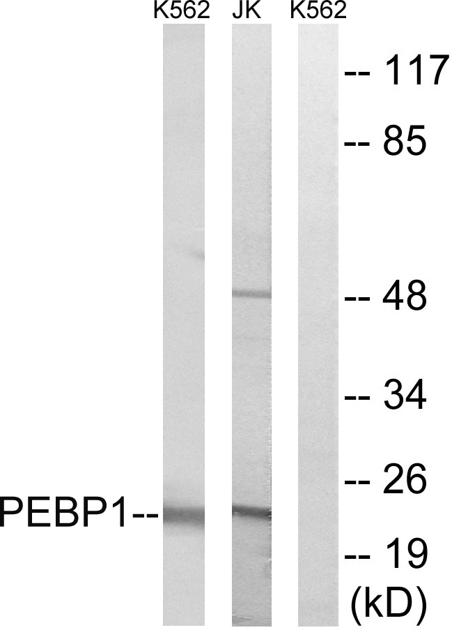

Anti-PEBP1 Antibody
A100812
ApplicationsWestern Blot, ELISA
Product group Antibodies
ReactivityHuman
Overview
- SupplierAntibodies.com
- Product NameAnti-PEBP1 Antibody
- Delivery Days Customer7
- ApplicationsWestern Blot, ELISA
- CertificationResearch Use Only
- ClonalityPolyclonal
- ConjugateUnconjugated
- HostRabbit
- IsotypeIgG
- Scientific DescriptionRabbit polyclonal antibody to PEBP1.
- ReactivityHuman
- UNSPSC12352203
Related products
Product group Antibodies
Anti-PEBP1 Antibody144-00578
ApplicationsImmunoFluorescence, ImmunoPrecipitation, Western Blot, Other Application
ReactivityHuman, Mouse
TargetPEBP1
- SizePrice

Product group Antibodies
Anti-PBP/PEBP1 Antibody Picoband(r)A01668-CARRIER-FREE
ApplicationsFlow Cytometry, Western Blot, ImmunoHistoChemistry
ReactivityHuman, Mouse
TargetPEBP1
- SizePrice
Product group Antibodies
PBP Recombinant AntibodyBSM-61243R
ApplicationsImmunoFluorescence, ImmunoPrecipitation, Western Blot, ImmunoCytoChemistry, ImmunoHistoChemistry, ImmunoHistoChemistry Frozen, ImmunoHistoChemistry Paraffin
TargetPEBP1
- SizePrice
Product group Antibodies
PEBP1 AntibodyCSB-PA004014
ApplicationsWestern Blot, ELISA
ReactivityHuman
TargetPEBP1
- SizePrice
Product group Antibodies
References

Goat anti-PEBP1EB05403
ApplicationsWestern Blot, ELISA, ImmunoHistoChemistry
ReactivityBovine, Canine, Human, Mouse, Rat
TargetPEBP1
- SizePrice
Product group Antibodies
PEBP1 Polyclonal AntibodyCAC14544
ApplicationsWestern Blot, ELISA, ImmunoHistoChemistry
ReactivityMouse
TargetPEBP1
- SizePrice
Product group Antibodies

PEBP1 / RKIP AntibodyLS-C400976
ApplicationsWestern Blot, ELISA, ImmunoHistoChemistry
ReactivityHuman, Mouse, Rat
TargetPEBP1
- SizePrice
![ICC/IF analysis of PFA-fixed PC-3M cells using GTX01163 RKIP antibody [SC58-09]. Green : primary antibody Blue : DAPI Permeabilization : 0.25% Triton X-100 in PBS](https://www.genetex.com/upload/website/prouct_img/normal/GTX01163/GTX01163_20200303_ICC-IF_642_w_23053121_823.webp)
Product group Antibodies
References
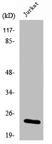
RKIP antibody [SC58-09]GTX01163
ApplicationsImmunoFluorescence, Western Blot, ImmunoCytoChemistry, ImmunoHistoChemistry, ImmunoHistoChemistry Paraffin
ReactivityHuman, Mouse
TargetPEBP1
- SizePrice

Product group Antibodies
Anti-PEBP1 AntibodyHPA008819
ApplicationsImmunoHistoChemistry
ReactivityHuman
TargetPEBP1
- SizePrice