

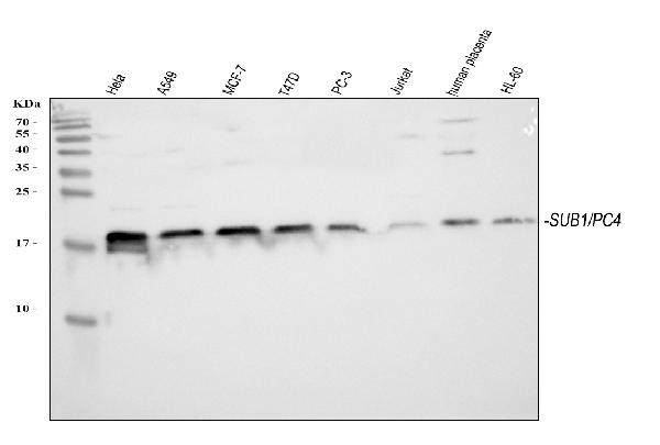
Figure 1. Western blot analysis of PC4/SUB1 using anti-PC4/SUB1 antibody (PB10098). Electrophoresis was performed on a 5-20% SDS-PAGE gel at 70V (Stacking gel) / 90V (Resolving gel) for 2-3 hours. The sample well of each lane was loaded with 30 ug of sample under reducing conditions. Lane 1: human Hela whole cell lysates, Lane 2: human A549 whole cell lysates, Lane 3: human MCF-7 whole cell lysates, Lane 4: human T47D whole cell lysates, Lane 5: human PC-3 whole cell lysates, Lane 6: human Jurkat whole cell lysates, Lane 7: human placenta tissue lysates, Lane 8: human HL-60 whole cell lysates. After electrophoresis, proteins were transferred to a nitrocellulose membrane at 150 mA for 50-90 minutes. Blocked the membrane with 5% non-fat milk/TBS for 1.5 hour at RT. The membrane was incubated with rabbit anti-PC4/SUB1 antigen affinity purified polyclonal antibody (Catalog # PB10098) at 0.5 microg/mL overnight at 4°C, then washed with TBS-0.1%Tween 3 times with 5 minutes each and probed with a goat anti-rabbit IgG-HRP secondary antibody at a dilution of 1:5000 for 1.5 hour at RT. The signal is developed using an Enhanced Chemiluminescent detection (ECL) kit (Catalog # EK1002) with Tanon 5200 system. A specific band was detected for PC4/SUB1 at approximately 19 kDa. The expected band size for PC4/SUB1 is at 14 kDa.
. Electrophoresis was performed on a 5-20% SDS-PAGE gel at 70V (Stacking gel) / 90V (Resolving gel) for 2-3 hours. The sample well of each lane was loaded with 30 ug of sample under reducing conditions. Lane 1: rat liver tissue lysates, Lane 2: rat spleen tissue lysates, Lane 3: rat stomach tissue lysates, Lane 4: rat RH35 whole cell lysates, Lane 5: mouse liver tissue lysates, Lane 6: mosue spleen tissue lysates. After electrophoresis, proteins were transferred to a nitrocellulose membrane at 150 mA for 50-90 minutes. Blocked the membrane with 5% non-fat milk/TBS for 1.5 hour at RT. The membrane was incubated with rabbit anti-PC4/SUB1 antigen affinity purified polyclonal antibody (Catalog # PB10098) at 0.5 microg/mL overnight at 4°C, then washed with TBS-0.1%Tween 3 times with 5 minutes each and probed with a goat anti-rabbit IgG-HRP secondary antibody at a dilution of 1:5000 for 1.5 hour at RT. The signal is developed using an Enhanced Chemiluminescent detection (ECL) kit (Catalog # EK1002) with Tanon 5200 system. A specific band was detected for PC4/SUB1 at approximately 19 kDa. The expected band size for PC4/SUB1 is at 14 kDa.")
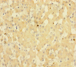
. PC4/SUB1 was detected in a paraffin-embedded section of human esophageal squamous carcinoma tissue. Heat mediated antigen retrieval was performed in EDTA buffer (pH 8.0, epitope retrieval solution). The tissue section was blocked with 10% goat serum. The tissue section was then incubated with 2 microg/ml rabbit anti-PC4/SUB1 Antibody (PB10098) overnight at 4°C. Peroxidase Conjugated Goat Anti-rabbit IgG was used as secondary antibody and incubated for 30 minutes at 37°C. The tissue section was developed using HRP Conjugated Rabbit IgG Super Vision Assay Kit (Catalog # SV0002) with DAB as the chromogen.")
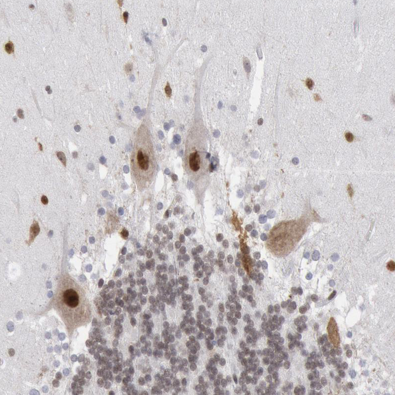
. PC4/SUB1 was detected in a paraffin-embedded section of human endometrioid adenocarcinoma type I tissue. Heat mediated antigen retrieval was performed in EDTA buffer (pH 8.0, epitope retrieval solution). The tissue section was blocked with 10% goat serum. The tissue section was then incubated with 2 microg/ml rabbit anti-PC4/SUB1 Antibody (PB10098) overnight at 4°C. Peroxidase Conjugated Goat Anti-rabbit IgG was used as secondary antibody and incubated for 30 minutes at 37°C. The tissue section was developed using HRP Conjugated Rabbit IgG Super Vision Assay Kit (Catalog # SV0002) with DAB as the chromogen.")
. PC4/SUB1 was detected in a paraffin-embedded section of human intestinal cancer tissue. Heat mediated antigen retrieval was performed in EDTA buffer (pH 8.0, epitope retrieval solution). The tissue section was blocked with 10% goat serum. The tissue section was then incubated with 5 microg/mL rabbit anti-PC4/SUB1 Antibody (PB10098) overnight at 4°C. DyLight488 Conjugated Goat Anti-Rabbit IgG (BA1127) was used as secondary antibody at 1:500 dilution and incubated for 30 minutes at 37°C. The section was counterstained with DAPI. Visualize using a fluorescence microscope and filter sets appropriate for the label used.")
Figure 1. Western blot analysis of PC4/SUB1 using anti-PC4/SUB1 antibody (PB10098). Electrophoresis was performed on a 5-20% SDS-PAGE gel at 70V (Stacking gel) / 90V (Resolving gel) for 2-3 hours. The sample well of each lane was loaded with 30 ug of sample under reducing conditions. Lane 1: human Hela whole cell lysates, Lane 2: human A549 whole cell lysates, Lane 3: human MCF-7 whole cell lysates, Lane 4: human T47D whole cell lysates, Lane 5: human PC-3 whole cell lysates, Lane 6: human Jurkat whole cell lysates, Lane 7: human placenta tissue lysates, Lane 8: human HL-60 whole cell lysates. After electrophoresis, proteins were transferred to a nitrocellulose membrane at 150 mA for 50-90 minutes. Blocked the membrane with 5% non-fat milk/TBS for 1.5 hour at RT. The membrane was incubated with rabbit anti-PC4/SUB1 antigen affinity purified polyclonal antibody (Catalog # PB10098) at 0.5 microg/mL overnight at 4°C, then washed with TBS-0.1%Tween 3 times with 5 minutes each and probed with a goat anti-rabbit IgG-HRP secondary antibody at a dilution of 1:5000 for 1.5 hour at RT. The signal is developed using an Enhanced Chemiluminescent detection (ECL) kit (Catalog # EK1002) with Tanon 5200 system. A specific band was detected for PC4/SUB1 at approximately 19 kDa. The expected band size for PC4/SUB1 is at 14 kDa.
Anti-PC4 Picoband Antibody
PB10098
ApplicationsImmunoFluorescence, Western Blot, ImmunoHistoChemistry
Product group Antibodies
ReactivityHamster, Human, Mouse, Rat
TargetSUB1
Overview
- SupplierBoster Bio
- Product NameAnti-PC4 Picoband Antibody
- Delivery Days Customer9
- Application Supplier NoteTested Species: In-house tested species with positive results. By Heat: Boiling the paraffin sections in 10mM citrate buffer, pH6.0, for 20mins is required for the staining of formalin/paraffin sections. Other applications have not been tested. Optimal dilutions should be determined by end users.
- ApplicationsImmunoFluorescence, Western Blot, ImmunoHistoChemistry
- CertificationResearch Use Only
- ClonalityPolyclonal
- Concentration500 ug/ml
- Gene ID10923
- Target nameSUB1
- Target descriptionSUB1 regulator of transcription
- Target synonymsP15, PC4, p14, activated RNA polymerase II transcriptional coactivator p15, SUB1 homolog, transcriptional regulator, activated RNA polymerase II transcription cofactor 4, positive cofactor 4
- HostRabbit
- IsotypeIgG
- Protein IDP53999
- Protein NameActivated RNA polymerase II transcriptional coactivator p15
- Scientific DescriptionBoster Bio Anti-PC4/SUB1 Antibody Picoband® catalog # PB10098. Tested in IF, IHC WB applications. This antibody reacts with Human, Mouse, Rat. The brand Picoband indicates this is a premium antibody that guarantees superior quality, high affinity, and strong signals with minimal background in Western blot applications. Only our best-performing antibodies are designated as Picoband, ensuring unmatched performance.
- ReactivityHamster, Human, Mouse, Rat
- Storage Instruction-20°C,2°C to 8°C
- UNSPSC12352203
Datasheet
MSDS
Related products
Product group Antibodies
Anti-SUB1 Antibody144-07070
ApplicationsImmunoFluorescence, ImmunoPrecipitation, Western Blot, ImmunoHistoChemistry
ReactivityHuman, Mouse
TargetSUB1
- SizePrice
Product group Antibodies
PC4 antibodyGTX65860
ApplicationsImmunoFluorescence, ImmunoPrecipitation, Western Blot, ImmunoCytoChemistry, ImmunoHistoChemistry, ImmunoHistoChemistry Paraffin
ReactivityHuman, Mouse
TargetSUB1
- SizePrice
Product group Antibodies
Anti-SUB1 AntibodyA31852
ApplicationsImmunoFluorescence, Western Blot, ImmunoHistoChemistry
ReactivityHuman, Mouse
- SizePrice

Product group Antibodies
Anti-PC4/SUB1 Antibody Picoband(r)A02698-1-CARRIER-FREE
ApplicationsImmunoFluorescence, Western Blot, ELISA, ImmunoHistoChemistry
ReactivityHuman, Mouse, Rat
TargetSUB1
- SizePrice
Product group Antibodies
SUB1 AntibodyCSB-PA022915HA01HU
ApplicationsELISA, ImmunoHistoChemistry
ReactivityHuman
TargetSUB1
- SizePrice
Product group Antibodies

SUB1 AntibodyLS-C346215
ApplicationsImmunoFluorescence, ImmunoPrecipitation, Western Blot, ImmunoHistoChemistry
ReactivityHuman, Mouse
TargetSUB1
- SizePrice
Product group Antibodies
Anti-SUB1 AntibodyHPA001311
ApplicationsWestern Blot, ImmunoCytoChemistry, ImmunoHistoChemistry
ReactivityHuman
TargetSUB1
- SizePrice