


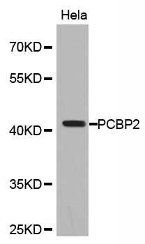
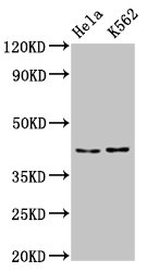
Figure 1. Western blot analysis of PCBP2/hnRNP E2 using anti-PCBP2/hnRNP E2 antibody (M02425). Electrophoresis was performed on a 5-20% SDS-PAGE gel at 70V (Stacking gel) / 90V (Resolving gel) for 2-3 hours. The sample well of each lane was loaded with 30 ug of sample under reducing conditions. Lane 1: human T-47D whole cell lysates, Lane 2: human HL-60 whole cell lysates, Lane 3: human A549 whole cell lysates, Lane 4: human HepG2 whole cell lysates, Lane 5: rat brain tissue lysates, Lane 6: mouse brain tissue lysates. After electrophoresis, proteins were transferred to a nitrocellulose membrane at 150 mA for 50-90 minutes. Blocked the membrane with 5% non-fat milk/TBS for 1.5 hour at RT. The membrane was incubated with mouse anti-PCBP2/hnRNP E2 antigen affinity purified monoclonal antibody (Catalog # M02425) at 0.5 microg/mL overnight at 4°C, then washed with TBS-0.1%Tween 3 times with 5 minutes each and probed with a goat anti-mouse IgG-HRP secondary antibody at a dilution of 1:10000 for 1.5 hour at RT. The signal is developed using an Enhanced Chemiluminescent detection (ECL) kit (Catalog # EK1001) with Tanon 5200 system. A specific band was detected for PCBP2/hnRNP E2 at approximately 39 kDa. The expected band size for PCBP2/hnRNP E2 is at 39 kDa.
. PCBP2/hnRNP E2 was detected in an immunocytochemical section of Caco-2 cells. Enzyme antigen retrieval was performed using IHC enzyme antigen retrieval reagent (AR0022) for 15 mins. The cells were blocked with 10% goat serum. And then incubated with 5 microg/mL mouse anti-PCBP2/hnRNP E2 Antibody (M02425) overnight at 4°C. DyLight®594 Conjugated Goat Anti-Mouse IgG (BA1141) was used as secondary antibody at 1:100 dilution and incubated for 30 minutes at 37°C. The section was counterstained with DAPI. Visualize using a fluorescence microscope and filter sets appropriate for the label used.")
. Overlay histogram showing PC-3 cells stained with M02425 (Blue line). To facilitate intracellular staining, cells were fixed with 4% paraformaldehyde and permeabilized with permeabilization buffer. The cells were blocked with 10% normal goat serum. And then incubated with mouse anti-PCBP2/hnRNP E2 Antibody (M02425, 1 microg/1x106 cells) for 30 min at 20°C. DyLight®488 conjugated goat anti-mouse IgG (BA1126, 5-10 microg/1x106 cells) was used as secondary antibody for 30 minutes at 20°C. Isotype control antibody (Green line) was mouse IgG (1 microg/1x106) used under the same conditions. Unlabelled sample without incubation with primary antibody and secondary antibody (Red line) was used as a blank control.")
Figure 1. Western blot analysis of PCBP2/hnRNP E2 using anti-PCBP2/hnRNP E2 antibody (M02425). Electrophoresis was performed on a 5-20% SDS-PAGE gel at 70V (Stacking gel) / 90V (Resolving gel) for 2-3 hours. The sample well of each lane was loaded with 30 ug of sample under reducing conditions. Lane 1: human T-47D whole cell lysates, Lane 2: human HL-60 whole cell lysates, Lane 3: human A549 whole cell lysates, Lane 4: human HepG2 whole cell lysates, Lane 5: rat brain tissue lysates, Lane 6: mouse brain tissue lysates. After electrophoresis, proteins were transferred to a nitrocellulose membrane at 150 mA for 50-90 minutes. Blocked the membrane with 5% non-fat milk/TBS for 1.5 hour at RT. The membrane was incubated with mouse anti-PCBP2/hnRNP E2 antigen affinity purified monoclonal antibody (Catalog # M02425) at 0.5 microg/mL overnight at 4°C, then washed with TBS-0.1%Tween 3 times with 5 minutes each and probed with a goat anti-mouse IgG-HRP secondary antibody at a dilution of 1:10000 for 1.5 hour at RT. The signal is developed using an Enhanced Chemiluminescent detection (ECL) kit (Catalog # EK1001) with Tanon 5200 system. A specific band was detected for PCBP2/hnRNP E2 at approximately 39 kDa. The expected band size for PCBP2/hnRNP E2 is at 39 kDa.
Anti-PCBP2/hnRNP E2 Antibody Picoband(r) (monoclonal, 4B9C7)
M02425-DYLIGHT488
ApplicationsFlow Cytometry, ImmunoFluorescence, Western Blot, ImmunoCytoChemistry
Product group Antibodies
ReactivityHuman, Mouse, Rat
TargetPCBP2
Overview
- SupplierBoster Bio
- Product NameAnti-PCBP2/hnRNP E2 Antibody Picoband(r) (monoclonal, 4B9C7)
- Delivery Days Customer9
- Application Supplier NoteTested Species: In-house tested species with positive results. Other applications have not been tested. Optimal dilutions should be determined by end users.
- ApplicationsFlow Cytometry, ImmunoFluorescence, Western Blot, ImmunoCytoChemistry
- CertificationResearch Use Only
- ClonalityMonoclonal
- Clone ID4B9C7
- Concentration500 ug/ml
- ConjugateDyLight 488
- Gene ID5094
- Target namePCBP2
- Target descriptionpoly(rC) binding protein 2
- Target synonymsHNRNPE2, HNRPE2, hnRNP-E2, poly(rC)-binding protein 2, alpha-CP2, epididymis secretory sperm binding protein, heterogeneous nuclear ribonucleoprotein E2, heterogenous nuclear ribonucleoprotein E2, hnRNP E2
- HostMouse
- IsotypeIgG2a
- Protein IDQ15366
- Protein NamePoly(rC)-binding protein 2
- Scientific DescriptionBoster Bio Anti-PCBP2/hnRNP E2 Antibody Picoband® (monoclonal, 4B9C7) catalog # M02425. Tested in Flow Cytometry, IF, ICC, WB applications. This antibody reacts with Human, Mouse, Rat. The brand Picoband indicates this is a premium antibody that guarantees superior quality, high affinity, and strong signals with minimal background in Western blot applications. Only our best-performing antibodies are designated as Picoband, ensuring unmatched performance.
- ReactivityHuman, Mouse, Rat
- Storage Instruction-20°C,2°C to 8°C
- UNSPSC12352203
Related products

Product group Antibodies
Anti-PCBP2/hnRNP E2 Antibody Picoband(r)A02425-2-CARRIER-FREE
ApplicationsFlow Cytometry, Western Blot, ELISA, ImmunoHistoChemistry
ReactivityHuman, Mouse, Rat
TargetPCBP2
- SizePrice
Product group Antibodies
PCBP2 Polyclonal AntibodyCAC15132
ApplicationsWestern Blot, ELISA, ImmunoHistoChemistry
TargetPCBP2
- SizePrice
Product group Antibodies
Anti-PCBP2 Antibody144-02531
ApplicationsImmunoFluorescence, Western Blot, ImmunoHistoChemistry
ReactivityHuman, Mouse, Rat
TargetPCBP2
- SizePrice
Product group Antibodies
PCBP2 antibody [C2C3], C-termGTX108362
ApplicationsImmunoFluorescence, Western Blot, ImmunoCytoChemistry
ReactivityHuman
TargetPCBP2
- SizePrice
Product group Antibodies
Anti-PCBP2 AntibodyA30576
ApplicationsImmunoFluorescence, Western Blot, ImmunoHistoChemistry
ReactivityHuman, Mouse, Rat
- SizePrice
Product group Antibodies
PCBP2 AntibodyCSB-PA622995LA01HU
ApplicationsWestern Blot, ELISA, ImmunoHistoChemistry
ReactivityHuman
TargetPCBP2
- SizePrice
Product group Antibodies
ApplicationsFlow Cytometry, Western Blot, ImmunoCytoChemistry
ReactivityHuman, Mouse, Rat
TargetPCBP2
- SizePrice

Product group Antibodies
Anti-PCBP2 AntibodyHPA038356
ApplicationsImmunoCytoChemistry, ImmunoHistoChemistry
ReactivityHuman
TargetPCBP2
- SizePrice